Showing posts with label Diseases. Show all posts

Biozorb Pros and Cons, Complications, Reviews

Biozorb is a major cancer treatment advancement. This novel technology was developed to enhance the precision and efficiency of radiotherapy for breast cancer patients. Biozorb is an innovative medical device with the potential to improve the accuracy of radiation therapy for breast cancer. The innovative design and features of the Biozorb have introduced a new era of cancer therapy as technology continues to transform healthcare.

What is Biozorb?

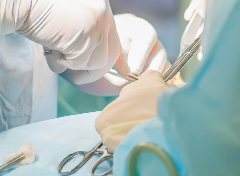
Biozorb is a bioabsorbable marker with a three-dimensional structure. During a lumpectomy, the surgeon places this device at the tumor site to assist the radiation therapy to be targeted correctly. The device improves cosmetic outcomes by supporting the breast tissue after surgery in addition to acting as a marker.
Biozorb Pros and Cons, Complications, Reviews


Pros 

Improved Radiation Targeting

Radiation targeting can be improved by Biozorb, which is one of its most important benefits. Oncologists may deliver radiation therapy with increased accuracy and lessen damage to the adjacent healthy tissues by offering a clear and exact image of the tumor spot.

Aids in Post-Treatment Monitoring

The distinctive structure of Biozorb aids in post-treatment monitoring. Using imaging methods like mammography, medical personnel can locate the device and track changes to make sure no abnormal tissue is growing there.

Better Cosmetic Results

Deformities can occasionally result after conventional breast cancer surgery. However, the support that Biozorb provides for breast tissue can help improve cosmetic results, giving the patient a more natural-looking appearance following treatment.

Radiation Exposure Reduced

Due to the precise localization of the tumor by Biozorb, radiation can be applied to the tumor only, sparing healthy tissues. For patients receiving radiation therapy, this may result in fewer adverse effects and an improvement in their quality of life.

Cons

Risks of Surgical Placement

Risks associated with the surgical installation of Biozorb include infection, bleeding, and pain. When evaluating the use of the device, surgeons must carefully balance these dangers against the potential advantages.

Imaging Errors

In some instances, the presence of Biozorb can produce artifacts on imaging scans, leading to possible confusion or misinterpretation of results. For precise evaluations, radiologists must be aware of these artifacts.

Limited Availability and Expertise

The surgical insertion of the specialized gadget Biozorb necessitates the appropriate training. Its availability may be restricted to medical centers with the required expertise, potentially limiting patient access.

Potential Complications

Every medical procedure has risks. Migration, tissue response, or discomfort with the device are all potential concerns with biozorb use. These possible problems must be explained to patients before therapy.

Complications

Surgical Risks

The implantation of Biozorb entails inherent risks, including bleeding, anesthesia issues, and scars. Patients must discuss these hazards with their surgeon prior to the procedure.

Infection Concerns

Any implant has the potential to raise concerns about infections at the surgical site. However, appropriate sterile techniques and postoperative care can substantially reduce this risk. The incision site should be closely monitored for indicators of infection, such as redness, swelling, and discharge.

Migration Issues

The Biozorb implants are intended to stay in place until they are absorbed, but there have been instances where migration has caused the device to move from its initial location. This might affect how precisely radiation therapy works. Migratory problems can be quickly identified and treated with close monitoring and follow-up with healthcare providers.

Reviews

According to a research article published on the NCBI website, the BioZorb arm exhibited considerable reductions in clinical target volume (CTV) and planned target volume (PTV), but not in ipsilateral lung or heart irradiation.

In 46% of cases, radiation oncologists considered BioZorb useful for reducing the boost planning target volume, according to a survey cited in an article published in the journal Anticancer Research.

Infantigo: Unraveling the Various Names of this Common Skin Condition

Infantigo is a common and contagious skin condition that affects both children and adults alike. It is known by several names, such as school sores, infintigo, infatigo, infentigo, enfantago, infantago, and infitigo. Despite the variety of names, it all refers to the same skin infection caused by bacteria. 

Infantigo is primarily caused by two types of bacteria: Streptococcus pyogenes and Staphylococcus aureus. It commonly appears as red sores around the mouth and nose, which can later burst, forming a yellowish-brown crust. Other symptoms may include itching, pain, and swelling in the affected area. Infantigo is also known by various other names such as:

infintigo, infatigo, infentigo, enfantago, infantago, and infitigo

School Sores

The term "school sores" is derived from the fact that infantigo is prevalent among school-going children due to their close contact in classrooms and playgrounds. The infection spreads easily through direct contact, leading to its association with educational institutions.

Infintigo

Infintigo is a misspelling of infantigo, but it is sometimes used interchangeably. Such misspellings might have occurred due to phonetic similarities or typographical errors.

Infatigo

Similar to infintigo, infatigo is another variant of infantigo that results from inadvertent misspellings. The presence of multiple names can sometimes lead to confusion, but they all refer to the same skin condition.

Infentigo

Infentigo is yet another name for infantigo, and it likely stems from regional or dialectal differences in pronunciation and spelling.

Enfantago

The term "enfantago" might have originated from languages where "e" is used as a prefix to indicate "in" or "on," thereby referring to a condition that affects infants. However, enfantago refers to the same condition as infantigo.

Infantago

Infantago is another variation of the Infantigo, probably influenced by regional linguistic nuances or colloquial usage. 

Infitigo

Infitigo is another term occasionally used for infantigo, potentially emerging from similar linguistic alterations.

Infintago

The name "infintago" is yet another variation of the term Infintago, reflecting the diversity of regional nomenclature for the same condition.

Origins of the Multiple Names

The various names of infantigo have emerged over time through a combination of typographical errors, regional language differences, and colloquial usages. As the condition spreads, different communities and cultures may adapt the name according to their local dialects, leading to the proliferation of alternative names.

Treatment and Prevention

Infantigo is usually not severe and can be treated with antibiotics prescribed by a healthcare professional. However, prevention is essential to avoid the spread of the infection. Simple hygiene practices, such as regular handwashing and avoiding contact with infected individuals, can significantly reduce the risk of contracting infantigo.

Equine Neurectomy Pros and Cons

Equine neurectomy is a surgical treatment that involves cutting or removing a particular nerve, frequently the palmar/plantar digital nerves, in order to treat horses' persistent leg or hoof pain. This procedure disrupts the nerve's capacity to convey pain signals to the brain, thereby lowering or eliminating pain feelings in the affected area.

Equine Neurectomy Pros and Cons

The horse is given general anesthesia throughout the neurectomy process to ensure its safety and well-being. The veterinarian uses modern imaging tools to identify the targeted nerve and makes a tiny incision around the nerve site. Depending on the procedure, the nerve is then cut off or eliminated. The incision is carefully stitched up, and the horse is allowed time to recover from anesthesia.

Pros

Pain Relief

Equine neurectomy's ability to significantly reduce the pain experienced by horses with chronic limb or hoof pain is its main advantage. By blocking the pain impulses, the horse feels a significant reduction in discomfort, resulting in a better quality of life.

Enhancing Performance

Following a neurectomy, horses whose performance had previously been hampered by chronic pain frequently display improved performance. They are able to move with greater freedom and perform better in a variety of sports, including racing, jumping, and dressage, when they are pain-free.

Management of Chronic Conditions

Equine neurectomy is a viable alternative to conventional therapies in the management of chronic problems like laminitis or navicular syndrome. When all other measures have failed, it provides an alternate strategy for easing pain.

Cons

Loss of Feeling

The fact that neurectomy permanently destroys sensation in the affected area is a big problem. While this can help with pain management, it may also conceal injuries or other problems that the horse might have had in a normal situation.

Limited diagnostic abilities

Once the nerve is cut off it is more difficult to diagnose any potential problems that might develop later on in the treated area. Future treatment approaches may become more complicated as veterinarians struggle to identify new diseases or the cause of discomfort.

Risk of Overuse and Dependency

In some situations, neurectomy may be considered a quick remedy to a problem, leading to its misuse. Without treating the underlying source of the pain, relying entirely on this technique could result in dependency and future consequences.

Considering the Ethics

It is important to not take the decision to perform an equine neurectomy lightly. It raises moral concerns about the well-being of the horse and the procedure's possible long-term effects. Before making a final decision, horse owners and veterinarians should carefully assess whether neurectomy is the best option and look into alternative therapies.

Equine Neurectomy Alternatives

Horse owners and veterinarians should look into less invasive options before choosing neurectomy:

Medications

Without resorting to surgery, a number of drugs can assist manage pain and inflammation. Non-steroidal anti-inflammatory medicines (NSAIDs) and other painkillers may be effective and should be taken into consideration.

Rehabilitation and physical therapy

Physical therapy and rehabilitation can be incorporated into the horse's routine to assist recovery and naturally reduce pain. Controlled exercise, cold therapy, and heat therapy are all effective methods.

Alternative Therapies

Alternative pain management methods could be investigated as a complementary therapy, including acupuncture, chiropractic adjustments, and herbal supplements.

Sclerosing Epithelioid Fibrosarcoma Symptoms, Causes, Diagnosis, Treatment

What is Sclerosing Epithelioid Fibrosarcoma?

Sclerosing epithelioid fibrosarcoma (SEF) is a rare type of cancer that arises from fibrous connective tissue. It was first described in 1995 and has since been classified as a distinct type of fibrosarcoma due to its unique histological characteristics.

SEF is most commonly found in young adults, with a slight predilection for females. It usually arises in the deep soft tissues of the extremities, but can also occur in the trunk, head and neck region. SEF is a slow-growing tumor that tends to infiltrate surrounding tissues, making complete surgical excision challenging.

SEF is typically diagnosed through a combination of clinical examination, radiographic imaging, and histopathological analysis of a biopsy specimen. The tumor has a distinct appearance on imaging studies, often showing a solid, well-circumscribed mass with areas of calcification. Histologically, SEF is characterized by small, uniform, round cells arranged in nests or cords within a hyalinized stroma.

Sclerosing Epithelioid Fibrosarcoma Symptoms, Causes, Diagnosis, Treatment

Symptoms

The symptoms of sclerosing epithelioid fibrosarcoma (SEF) can vary depending on the location and size of the tumor. In some cases, SEF may not cause any symptoms until it grows to a size that compresses or invades nearby structures.

Common symptoms of SEF may include:

  • A painless lump or swelling in the affected area
  • Limited range of motion or stiffness in nearby joints
  • Numbness or tingling in the affected limb or body part
  • Weakness or loss of function in the affected limb or body part
  • Persistent pain that may worsen with activity or movement
  • Weight loss, fatigue, or other non-specific symptoms in advanced cases

Causes

The exact cause of sclerosing epithelioid fibrosarcoma (SEF) is not well understood. However, it is believed to arise from a genetic mutation or abnormality that leads to the uncontrolled growth of cells in fibrous connective tissue.

There is evidence to suggest that SEF may be associated with a genetic disorder called familial adenomatous polyposis (FAP), which is an inherited condition that increases the risk of developing polyps in the colon and rectum. Studies have found that individuals with FAP have an increased risk of developing SEF, particularly in the soft tissues of the extremities.

Other factors that may contribute to the development of SEF include exposure to radiation or certain chemicals, as well as trauma or injury to the affected area. However, the role of these factors in the development of SEF is not well established and more research is needed to understand the underlying causes of this rare cancer.

Diagnosis

Diagnosing sclerosing epithelioid fibrosarcoma (SEF) typically involves a combination of clinical examination, radiographic imaging, and histopathological analysis of a biopsy specimen.

The diagnostic process for SEF may involve the following steps:

Medical history and physical exam

A healthcare provider will review the patient's medical history and perform a physical exam to assess the location, size, and characteristics of the tumor.

Imaging tests

Imaging tests such as X-rays, CT scans, MRI scans, or PET scans may be used to visualize the tumor and evaluate its extent and location.

Biopsy

A biopsy is typically needed to confirm the diagnosis of SEF. During a biopsy, a small sample of the tumor tissue is removed and examined under a microscope by a pathologist to identify the presence of characteristic features of SEF.

Immunohistochemistry

Immunohistochemistry is a laboratory technique that uses specific antibodies to detect proteins or other markers in a tissue sample. It can help confirm the diagnosis of SEF and distinguish it from other types of tumors that may have similar characteristics.

Treatment

The treatment of sclerosing epithelioid fibrosarcoma (SEF) typically involves a multimodal approach that includes surgical excision, radiation therapy, and chemotherapy. The specific treatment plan will depend on factors such as the size, location, and extent of the tumor, as well as the patient's overall health and individual preferences.

Surgery

Surgery is the mainstay of treatment for SEF and involves the complete removal of the tumor. However, complete surgical excision of SEF can be challenging due to the tumor's tendency to infiltrate surrounding tissues. In some cases, amputation or limb-sparing surgery may be necessary to achieve complete removal of the tumor.

Radiation therapy

Radiation therapy may be used in combination with surgery to treat SEF. It involves the use of high-energy radiation to kill cancer cells and shrink the tumor. Radiation therapy may also be used as a palliative treatment to relieve pain and other symptoms in advanced cases.

Chemotherapy

Chemotherapy may be used to treat SEF that has spread to other parts of the body. However, SEF is typically resistant to chemotherapy, and its role in the treatment of this cancer is not well established.

Clinical trials

There is ongoing research into new treatments for SEF, and patients may be eligible to participate in clinical trials that test new drugs or therapies.

What is primordial cyst? - Location, Causes, Treatment

 What is primordial cyst?

A primordial cyst, also known as a dentigerous cyst or follicular cyst, is a type of odontogenic cyst that can develop in the jaws. These cysts usually arise from the follicle that surrounds the crown of an unerupted tooth, or from remnants of the dental lamina (an embryonic structure that gives rise to teeth). Primordial cysts are often asymptomatic and may be discovered incidentally on dental radiographs. However, in some cases, they can cause pain, swelling, or displacement of adjacent teeth.

Location

Primordial cysts can occur in either the maxilla (upper jaw) or the mandible (lower jaw), and are typically found near the crown of an unerupted or impacted tooth. The most common sites for these cysts are the mandibular third molars (wisdom teeth) and the maxillary canines. However, they can also occur in association with other teeth and in other locations in the jaw.

What is primordial cyst - Location, Causes, Treatment

Causes

The exact cause of primordial cysts is not fully understood, but it is believed to result from the accumulation of fluid between the reduced enamel epithelium and the enamel of an unerupted or impacted tooth. This fluid buildup can cause pressure to build up, leading to the formation of a cyst.

In some cases, a primordial cyst may develop from remnants of the dental lamina, which is an embryonic structure that gives rise to teeth.

Certain factors may increase the risk of developing a primordial cyst, such as poor oral hygiene, trauma to the jaw, and genetic predisposition. However, in many cases, the development of these cysts is spontaneous and not related to any specific risk factors.

Treatment

The treatment for a primordial cyst depends on the size and location of the cyst, as well as the patient's symptoms and overall health. In many cases, if the cyst is small and asymptomatic, it may not require immediate treatment, but should be monitored regularly by a dentist or oral surgeon to ensure that it does not grow or cause any problems.

If the cyst is larger or causing discomfort, treatment may involve surgical removal. The procedure typically involves making an incision in the gum tissue to access the cyst and removing it, along with any associated tooth, if necessary. In some cases, a small drain may be placed to promote healing and prevent the buildup of fluid.

After the cyst is removed, the patient may be prescribed pain medication and antibiotics to prevent infection. It is important to maintain good oral hygiene after the procedure and follow any post-operative instructions provided by the dentist or oral surgeon.

Regular follow-up appointments will be necessary to ensure that the cyst does not recur or cause any further problems.

Optic disc cupping Meaning, Symptoms, Causes, Treatment | Optic disc cupping vs Normal

Optic disc cupping Meaning

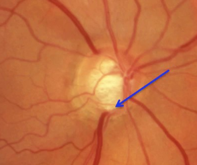
Optic disc cupping is a condition that occurs when the optic nerve head, which is the point where the optic nerve enters the eye, appears to have a depression or a concave shape. This depression is called the cup and is a normal feature of the optic disc. However, when the cup becomes larger, it can indicate the development of certain eye diseases, such as glaucoma.

In glaucoma, the pressure inside the eye increases, which can damage the optic nerve and lead to vision loss. As the damage progresses, the cup becomes larger and deeper. Therefore, the measurement of the cup-to-disc ratio is an important diagnostic tool for assessing the severity of glaucoma and monitoring its progression.

Optic disc cupping Symptoms

Optic disc cupping itself does not cause any symptoms. In fact, it is often detected during a routine eye exam by an ophthalmologist or optometrist who is looking for signs of eye disease.

However, optic disc cupping can be a sign of an underlying eye condition, such as glaucoma. Glaucoma is a progressive eye disease that can cause damage to the optic nerve and lead to vision loss if left untreated. Symptoms of glaucoma may include:

  • Blurred or hazy vision
  • Loss of peripheral vision (side vision)
  • Difficulty adjusting to dark rooms
  • Halos around lights
  • Eye pain or redness
Optic disc cupping Meaning, Symptoms, Causes, Treatment  Optic disc cupping vs Normal

Optic disc cupping Causes

Optic disc cupping can be caused by several underlying eye conditions, including:

Glaucoma

The most common cause of optic disc cupping is glaucoma, a group of eye diseases that damages the optic nerve, often due to elevated pressure within the eye. As glaucoma progresses, the cup-to-disc ratio increases, and the cup becomes deeper and wider.

Optic neuropathy

Optic neuropathy refers to any damage to the optic nerve that impairs its function. This condition can cause optic disc cupping, among other symptoms.

Optic nerve head drusen

Optic nerve head drusen is a condition in which small calcified deposits accumulate in the optic nerve head. This can lead to a small or large cup-to-disc ratio, depending on the number and location of the deposits.

Optic disc edema

Optic disc edema is a swelling of the optic disc, often caused by increased pressure within the skull or inflammation in the eye. This condition can cause a shallow cup-to-disc ratio.

Other conditions

Rarely, other conditions such as retinal detachment, retinal vein occlusion, and optic neuritis can cause optic disc cupping.

Optic disc cupping Treatment

The treatment for optic disc cupping depends on the underlying condition causing the cupping. Here are some common treatments for the most common cause of optic disc cupping, glaucoma:

Medications

Eye drops or oral medications can be prescribed to lower intraocular pressure (IOP), which can slow or halt the progression of glaucoma.

Laser therapy

Laser trabeculoplasty is a type of laser therapy that can be used to open up drainage channels in the eye, which can help lower IOP.

Surgery

Trabeculectomy is a surgical procedure that can help lower IOP by creating a new drainage channel in the eye.

Regular eye exams

Regular eye exams are important for monitoring the progression of optic disc cupping and glaucoma, and adjusting treatment as needed.

Optic disc cupping vs Normal

The optic disc is the part of the retina where the optic nerve exits the eye. In a normal optic disc, there is a central area called the "optic cup," which is a small depression in the center of the disc. In a healthy eye, the size of the optic cup is smaller than the size of the optic disc.

In contrast, in optic disc cupping, the size of the optic cup is enlarged and the edges of the cup become more pronounced, while the optic disc itself may appear thinner. Optic disc cupping is a sign of damage to the optic nerve and can be caused by several conditions, including glaucoma, optic neuropathy, and optic nerve head drusen.

What are Tardieu Spots? - Definition, Location, Causes | Tardieu spots vs Petechiae

What are Tardieu spots?

Tardieu spots, also known as petechiae Tardieu or Tardieu's points, are small, red or purple spots that can appear on the skin after death. These spots are caused by the accumulation of blood in the small blood vessels under the skin, which occurs as a result of gravity and the pooling of blood in the lowest parts of the body.

Tardieu spots are often used as a forensic indicator to estimate the time of death, as their presence and distribution on the body can provide clues about how long a person has been deceased. However, it is important to note that other factors, such as medical conditions and injuries, can also affect the development and distribution of Tardieu spots. Therefore, their presence or absence should be interpreted in conjunction with other evidence and forensic analysis.

Tardieu Spots Definition

Tardieu spots, also known as petechiae Tardieu or Tardieu's points, are small, reddish-purple spots or bruises that can appear on the skin after death. These spots are caused by the accumulation of blood in the small blood vessels under the skin due to gravity and the pooling of blood in the lowest parts of the body. Tardieu spots are often used in forensic investigations to help estimate the time of death, as their presence and distribution on the body can provide clues about how long a person has been deceased. However, other factors can also affect the development and distribution of Tardieu spots, so their presence or absence should be interpreted in conjunction with other evidence and forensic analysis.

Tardieu Spots Location

Tardieu spots can appear in various locations on the body, but they tend to be most common in areas where blood vessels are close to the skin's surface. These areas include the neck, face, eyelids, chest, abdomen, and inner surfaces of the arms and legs.

The distribution of Tardieu spots on the body can provide important information for forensic investigators trying to estimate the time of death. For example, if the spots are concentrated in areas that are typically in contact with a surface, such as the face or chest, this may indicate that the body was in that position for an extended period after death. Conversely, if the spots are more evenly distributed across the body, this may suggest that the body was moved or manipulated soon after death. However, other factors such as medical conditions and injuries can also affect the distribution of Tardieu spots, so their interpretation should always be done in conjunction with other forensic evidence.

Tardieu Spots Causes

Tardieu spots are caused by the accumulation of blood in the small blood vessels under the skin after death. This occurs due to the gravitational effect on the blood and the pooling of blood in the lowest parts of the body. The pressure of the accumulating blood causes the vessels to rupture, leading to the formation of small reddish-purple spots or bruises on the skin.

The accumulation of blood can be influenced by various factors, including the position of the body after death, the time elapsed since death, and the presence of underlying medical conditions that affect blood clotting or vessel integrity. For example, if the body is lying in a face-down position, Tardieu spots may be more prominent on the front of the body. If the body has been dead for a longer period, Tardieu spots may be more widespread and numerous. Medical conditions such as bleeding disorders or sepsis can also increase the likelihood and severity of Tardieu spots.

Overall, Tardieu spots are a postmortem phenomenon that can provide important clues for forensic investigators about the circumstances surrounding a person's death. However, their interpretation must always be considered in conjunction with other forensic evidence to reach a definitive conclusion.

Tardieu Spots vs Petechiae

Tardieu spots and petechiae are similar in appearance, as they both present as small, reddish-purple spots on the skin. However, they have different underlying causes and can appear in different contexts.

Tardieu spots are a postmortem phenomenon that occurs due to the accumulation of blood in the small blood vessels under the skin after death. The pressure of the accumulating blood causes the vessels to rupture, leading to the formation of small reddish-purple spots on the skin. Tardieu spots are commonly used as a forensic indicator to estimate the time of death.

Petechiae, on the other hand, are caused by the rupture of small blood vessels under the skin due to increased pressure or trauma. Petechiae can occur in various medical conditions, including bleeding disorders, infections, and certain medications. Petechiae can also occur in non-medical contexts, such as in cases of strangulation, where pressure on the neck can cause the rupture of blood vessels and the appearance of petechiae.

IHSS heart Meaning, Symptoms, Causes, Treatment

IHSS heart Meaning

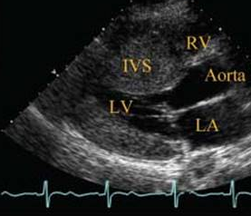
Idiopathic hypertrophic subaortic stenosis (IHSS), also known as hypertrophic obstructive cardiomyopathy (HOCM), is a medical condition in which the heart muscle becomes abnormally thickened (hypertrophied), especially in the area of the left ventricle, which is the chamber of the heart responsible for pumping blood to the body. This thickening can cause narrowing (stenosis) of the pathway for blood to leave the heart and can also cause problems with the heart's ability to relax and fill with blood.

IHSS heart Symptoms

The symptoms of idiopathic hypertrophic subaortic stenosis (IHSS) can vary depending on the severity of the condition, but common symptoms may include:

  • Shortness of breath, especially during exercise or physical activity
  • Chest pain, discomfort or pressure
  • Fainting, dizziness, or lightheadedness, particularly with exertion or after standing up
  • Palpitations or rapid heartbeat
  • Fatigue or weakness
  • Difficulty breathing while lying flat
IHSS heart Meaning, Symptoms, Causes, Treatment

IHSS heart Causes

The exact cause of idiopathic hypertrophic subaortic stenosis (IHSS) is not fully understood. However, there are several factors that may contribute to the development of the condition, including:

Genetic mutations

In some cases, IHSS may be inherited in an autosomal dominant pattern, meaning that a person only needs to inherit one copy of the mutated gene from one parent to develop the condition.

Abnormal protein production

People with IHSS may have abnormal protein production in the heart muscle, leading to excessive thickening of the heart muscle.

High blood pressure

Chronic high blood pressure can cause the heart muscle to thicken and become stiff, which may increase the risk of developing IHSS.

Aging

As people age, the heart muscle may naturally thicken, which can increase the risk of developing IHSS.

Unknown factors

In some cases, the cause of IHSS may be unknown (idiopathic).

IHSS heart Meaning Treatment

The treatment for idiopathic hypertrophic subaortic stenosis (IHSS) depends on the severity of the condition, the presence and severity of symptoms, and the overall health of the affected person. Treatment options may include:

Medications

Medications such as beta-blockers, calcium channel blockers, and antiarrhythmic drugs may be prescribed to help reduce symptoms, control heart rate, and prevent dangerous heart rhythms.

Surgical procedures

Surgical procedures such as septal myectomy or alcohol septal ablation may be performed to remove part of the thickened heart muscle and widen the pathway for blood to leave the heart.

Implantable devices

In some cases, an implantable cardioverter-defibrillator (ICD) or pacemaker may be implanted to help regulate the heart's rhythm and prevent dangerous heart rhythms.

Lifestyle modifications

Lifestyle modifications such as avoiding strenuous physical activity, managing stress, and avoiding certain medications may be recommended to help reduce symptoms and improve overall heart health.

What is Air Enema? - Procedure, Complications | Air enema vs Barium enema

What is Air Enema? 

An air enema is a medical procedure in which air is introduced into the rectum and colon through the anus, typically using a specialized enema apparatus. The purpose of an air enema is to help diagnose and treat certain medical conditions, such as blockages or obstructions in the bowel.

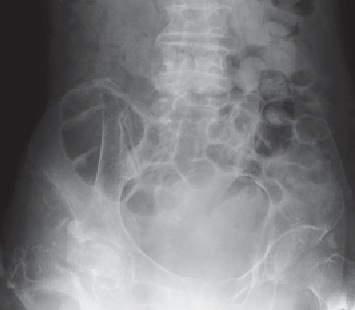
During the procedure, the patient lies on their back while a small tube is inserted into the rectum. Air is then slowly introduced into the colon, causing it to expand and allowing any obstructions or blockages to be visualized on X-rays or other imaging studies.

Air enemas can also be used therapeutically to relieve constipation or to help prepare the bowel for certain medical procedures, such as a colonoscopy. However, the procedure can cause discomfort or pain, and there is a risk of complications such as perforation of the bowel, so it is typically only used when other diagnostic or therapeutic options have been exhausted or are not suitable.

What is Air Enema? - Procedure, Complications | Air enema vs Barium enema

Procedure

An air enema is a medical procedure that is typically performed in a hospital or clinic setting by a healthcare professional, such as a radiologist or gastroenterologist. Here is a general overview of the steps involved in an air enema procedure:

Preparation

Before the procedure, the patient may be asked to refrain from eating or drinking for a certain period of time to help ensure an empty bowel. The patient may also be given medication to help relax or sedate them, depending on the circumstances.

Positioning

The patient is typically positioned on their back on an X-ray table, with their knees bent and their feet resting in stirrups to expose the anus.

Insertion of the enema tube

A lubricated tube is gently inserted into the rectum through the anus. The tube may be equipped with a balloon at the end, which is inflated to help keep the tube in place.

Introduction of air

Once the tube is in place, air is slowly introduced into the colon using a specialized device attached to the enema tube. The rate and volume of air introduced may be adjusted based on the patient's response and the desired outcome.

Monitoring and imaging

During the procedure, the healthcare professional may use imaging techniques such as X-rays or ultrasound to monitor the expansion of the colon and look for any signs of obstructions or blockages.

Completion and aftercare

Once the procedure is complete, the tube is removed and the patient may be given instructions for aftercare, such as drinking fluids or taking laxatives to help clear the colon. The healthcare professional will also monitor the patient for any signs of complications, such as pain or bleeding.

Complications

While an air enema is generally considered safe, there are some potential complications that can occur, including:

Bowel perforation

If too much air is introduced or if the colon is already weakened or damaged, the pressure from the air can cause a tear or perforation in the bowel.

Bleeding

The insertion of the enema tube or the pressure from the air can cause small tears in the rectum or colon, leading to bleeding.

Infection

There is a risk of infection from the insertion of the enema tube or from the air itself, particularly if there is an existing bowel infection or inflammation.

Pain or discomfort

The introduction of air into the colon can cause cramping, discomfort, or pain. This may be particularly true if there is an underlying condition such as irritable bowel syndrome (IBS).

Electrolyte imbalance

An air enema may cause a shift in electrolyte levels in the body, which can lead to complications such as dehydration or abnormal heart rhythms.

Air enema vs Barium enema 

Air enema and barium enema are two medical procedures that are used to diagnose and treat certain conditions affecting the colon and rectum. While both procedures are designed to help visualize the colon and rectum on imaging studies, they differ in several ways.

The primary difference between the two procedures is the substance used. In an air enema, air is introduced into the colon to help visualize any blockages or obstructions. In contrast, a barium enema involves introducing a liquid solution containing barium sulfate into the colon. Barium sulfate shows up clearly on X-rays, allowing healthcare professionals to visualize the colon and rectum more clearly.

Another difference is in the procedure and preparation. An air enema typically involves the insertion of a small tube into the rectum through the anus, while a barium enema involves a larger tube being inserted into the rectum. The preparation for each procedure may also differ, with a barium enema often requiring more extensive bowel preparation, such as the use of laxatives or enemas.

Rectosigmoid Junction Location, Polyp, Cancer, Surgery

The rectosigmoid junction is a part of the large intestine that connects the sigmoid colon to the rectum. It is located in the lower left quadrant of the abdomen, near the pelvis. The sigmoid colon is a curved section of the large intestine that connects the descending colon to the rectosigmoid junction. The rectum is the final segment of the large intestine that connects to the anus. The rectosigmoid junction plays an important role in the elimination of waste products from the body. As stool passes through the sigmoid colon, it is gradually dehydrated and compacted, forming solid feces. When the feces reach the rectosigmoid junction, the muscles of the colon contract, propelling the feces into the rectum and triggering the urge to defecate.

Function

The rectosigmoid junction is a crucial part of the large intestine that performs several important functions in the digestive process. Its main functions are:

Storage of feces

The rectosigmoid junction serves as a temporary storage site for feces before they are eliminated from the body. The muscles of the rectum and anus are designed to hold fecal matter until a suitable time and place for defecation.

Formation of solid feces

The rectosigmoid junction plays a key role in the formation of solid feces from the liquid waste material that enters the large intestine. As the feces pass through the sigmoid colon, they are gradually dehydrated and compacted, forming a more solid consistency.

Elimination of waste products

The rectosigmoid junction is responsible for propelling fecal matter from the large intestine into the rectum and triggering the urge to defecate. This process is initiated by the contraction of the muscles of the colon and rectum.

Absorption of water and electrolytes

The rectosigmoid junction absorbs water and electrolytes from the fecal matter, which helps to regulate the balance of fluids and electrolytes in the body.

Rectosigmoid Junction Location, Polyp, Cancer, Surgery

Location

The rectosigmoid junction is located in the lower left quadrant of the abdomen, close to the pelvis. It is situated between the sigmoid colon, which is the S-shaped part of the large intestine, and the rectum, which is the final segment of the large intestine before the anus. The rectosigmoid junction is approximately 15-20 cm (6-8 inches) long and is located at the level of the third sacral vertebra, which is near the bottom of the spine.

Polyp

A rectosigmoid junction polyp is a growth that arises from the lining of the rectosigmoid junction, which is the area where the sigmoid colon connects to the rectum. These types of polyps are typically found during a routine colonoscopy, which is a procedure used to examine the colon and rectum for abnormalities.

Rectosigmoid junction polyps can be either benign or malignant, and their size and shape can vary. Some polyps are small and have a stalk-like base, while others are larger and have a broader base. Most polyps do not cause any symptoms, but some larger ones can cause bleeding or obstruct the bowel.

If a rectosigmoid junction polyp is found during a colonoscopy, it is typically removed during the same procedure. This is done using a specialized tool that is passed through the colonoscope. The polyp is either snared or cut off and then sent to a laboratory for analysis.

Cancer

Rectosigmoid junction cancer is a type of cancer that originates in the rectosigmoid junction, which is the area where the sigmoid colon connects to the rectum. It is a subtype of colorectal cancer, which is the third most common cancer diagnosed in both men and women.

Rectosigmoid junction cancer usually develops slowly over many years, starting as a small polyp or growth on the inner lining of the colon or rectum. Over time, the cells in the polyp may become cancerous and begin to grow and spread. The most common symptoms of rectosigmoid junction cancer include abdominal pain, rectal bleeding, changes in bowel habits, and unexplained weight loss.

The diagnosis of rectosigmoid junction cancer is typically made through a combination of medical imaging tests, such as CT scans or MRIs, and biopsies of the affected tissue. Treatment for rectosigmoid junction cancer usually involves a combination of surgery, chemotherapy, and radiation therapy. The specific treatment plan depends on the stage and location of the cancer, as well as the overall health of the patient.

The best way to prevent rectosigmoid junction cancer is through regular screening for colorectal cancer, which can detect and remove precancerous polyps before they become cancerous. The American Cancer Society recommends that adults at average risk for colorectal cancer begin screening at age 45, while those at higher risk may need to start screening at an earlier age.

Surgery

Surgery is often a primary treatment for rectosigmoid junction cancer, especially for early-stage cancers that have not spread to other parts of the body. The goal of surgery is to remove the cancerous tissue while preserving as much of the normal surrounding tissue as possible.

The type of surgery used for rectosigmoid junction cancer depends on the size and location of the tumor. In some cases, a minimally invasive procedure called a laparoscopic or robotic-assisted surgery may be used. This involves making small incisions in the abdomen and using specialized instruments and a camera to remove the tumor.

In more advanced cases, a more extensive surgery may be required to remove the rectum and sigmoid colon. This is called a low anterior resection or an abdominal perineal resection, depending on the location and extent of the cancer. During this procedure, the affected portion of the colon and rectum is removed, and the remaining ends of the colon and rectum are reconnected.

After surgery, patients may require additional treatment, such as chemotherapy or radiation therapy, to kill any remaining cancer cells and prevent the cancer from recurring. The specific treatment plan depends on the stage and location of the cancer, as well as the patient's overall health and other medical conditions.

Calyceal Diverticulum Symptoms, Causes, Ultrasound, Treatment

 A calyceal diverticulum is a rare urological condition that affects the kidneys. It is a small sac-like structure that forms in one of the calyces of the kidney, which is a structure that collects urine before it flows into the renal pelvis and ureter. Calyceal diverticula are usually benign and do not cause any symptoms, but they can sometimes cause urinary tract infections or kidney stones.

The exact cause of calyceal diverticula is unknown, but they are thought to be congenital, meaning they are present from birth. They can also be acquired, meaning they develop later in life as a result of kidney damage or infection. Calyceal diverticula are more common in women than men and typically occur in people between the ages of 30 and 50.

Calyceal Diverticulum Symptoms

Many people with calyceal diverticulum do not experience any symptoms and the condition may be discovered incidentally during a routine medical exam or imaging study. However, in some cases, calyceal diverticulum can cause symptoms such as:

Recurrent urinary tract infections

The diverticulum can become a breeding ground for bacteria, which can lead to recurrent infections.

Flank pain

In some cases, calyceal diverticulum may cause pain in the back or side of the affected kidney.

Hematuria

Blood in the urine may occur due to irritation of the lining of the diverticulum.

Kidney stones

Stones may form within the diverticulum or may develop in the urinary tract as a result of the diverticulum's presence.

Hydronephrosis

This condition occurs when urine builds up in the kidney and causes it to swell. It can occur if the diverticulum obstructs the flow of urine from the kidney.
Calyceal Diverticulum Symptoms, Causes, Ultrasound, Treatment

Calyceal Diverticulum Causes

The exact cause of calyceal diverticulum is not fully understood, but there are several theories that attempt to explain its development. Some possible causes of calyceal diverticulum are:

Congenital

Calyceal diverticulum may be present from birth and develop as a result of abnormal embryonic development.

Acquired

The condition may also develop later in life due to damage or inflammation of the kidney or urinary tract.

Obstruction

Obstruction of the calyceal system may result in an increase in pressure, which can cause the formation of a diverticulum.

Infection

In some cases, calyceal diverticulum may be associated with chronic urinary tract infections or pyelonephritis.

Trauma

Injury to the kidney or urinary tract may cause the development of a diverticulum.

Genetics

There may be a genetic predisposition to the development of calyceal diverticulum.

Calyceal Diverticulum Ultrasound

On an ultrasound image, a calyceal diverticulum may appear as a small, fluid-filled sac that protrudes from the calyx of the kidney. However, other conditions such as cysts or tumors may appear similar on ultrasound, and further imaging tests may be necessary to confirm a diagnosis.

Ultrasound is a safe and painless procedure that does not use radiation, making it a good option for patients who cannot have other imaging tests such as CT or MRI due to radiation exposure or other factors. However, the accuracy of ultrasound for diagnosing calyceal diverticulum depends on the size and location of the diverticulum, and it may not be able to detect small or deep-seated diverticula.

Calyceal Diverticulum Treatment

The treatment for calyceal diverticulum depends on the severity of the condition and the presence of symptoms. In many cases, no treatment is necessary if the diverticulum is small and does not cause any symptoms. However, if the diverticulum causes recurrent infections or kidney stones, treatment may be required.

Antibiotics

If the patient has a urinary tract infection, antibiotics may be prescribed to treat the infection and prevent future infections.

Pain relief

Over-the-counter pain relievers such as acetaminophen or ibuprofen may be used to relieve mild pain or discomfort.

Surgical intervention

If the diverticulum is large, deep-seated, or causing significant symptoms, surgery may be necessary. The type of surgery will depend on the size and location of the diverticulum and the overall health of the patient. Surgical options may include open surgery or minimally invasive techniques such as laparoscopy or robotic-assisted surgery.

Observation

In some cases, observation may be recommended to monitor the diverticulum for any changes or progression. Regular imaging tests may be necessary to monitor the condition.

Hypofibrinogenemia Definition, Symptoms, Causes, Retraction Time, Treatment

Hypofibrinogenemia is a medical condition characterized by low levels of fibrinogen in the blood. Fibrinogen is a protein that is essential for blood clotting, and its deficiency can result in excessive bleeding or hemorrhage. Hypofibrinogenemia can be inherited, acquired, or a combination of both.

Inherited hypofibrinogenemia is a rare genetic disorder that is caused by mutations in the genes that code for fibrinogen. It can be present at birth or may become apparent later in life.

Acquired hypofibrinogenemia can be caused by a variety of conditions, including liver disease, severe infections, disseminated intravascular coagulation (DIC), and certain medications. In some cases, hypofibrinogenemia may be a result of surgery, particularly in patients who have undergone extensive liver or cardiac surgery.

Hypofibrinogenemia Definition

Hypofibrinogenemia is a medical condition characterized by abnormally low levels of fibrinogen in the blood, which is a protein that plays a crucial role in blood clotting. This condition can be inherited or acquired and may result in excessive bleeding or hemorrhage. 

Hypofibrinogenemia Definition, Symptoms, Causes, Retraction Time, Treatment

Hypofibrinogenemia Symptoms

The symptoms of hypofibrinogenemia may vary depending on the severity of the condition and the underlying cause. Some common symptoms of hypofibrinogenemia include:

  • Prolonged bleeding after an injury or surgical procedure
  • Easy bruising
  • Frequent nosebleeds
  • Heavy menstrual bleeding
  • Bleeding gums
  • Blood in the urine or stool
  • Excessive bleeding during or after childbirth

Hypofibrinogenemia Causes

Hypofibrinogenemia can be caused by a variety of factors, including genetic mutations, underlying medical conditions, and certain medications. Some common causes of hypofibrinogenemia include:

Inherited hypofibrinogenemia

This is a rare genetic disorder that is caused by mutations in the genes that produce fibrinogen. Inherited hypofibrinogenemia can be present at birth or may develop later in life.

Liver disease

Fibrinogen is produced in the liver, so any liver disease that impairs liver function can lead to hypofibrinogenemia.

Disseminated intravascular coagulation

DIC is a medical condition that can result from severe infections, trauma, or other underlying medical conditions. It can cause clotting in small blood vessels throughout the body, which can deplete fibrinogen levels and result in hypofibrinogenemia.

Medications

Certain medications, such as fibrinolytic drugs used to treat blood clots, can reduce fibrinogen levels.

Surgery

Hypofibrinogenemia can occur as a complication of surgery, particularly in patients who have undergone extensive liver or cardiac surgery.

Other medical conditions

Hypofibrinogenemia can also occur as a result of other medical conditions, such as leukemia, lymphoma, or multiple myeloma.

Hypofibrinogenemia Retraction Time

Retraction time is a laboratory test that measures the time it takes for a blood clot to form and then retract or dissolve. The test is performed by making a small puncture in the skin and then applying a standard amount of pressure to the area to induce bleeding. A stopwatch is used to time how long it takes for the bleeding to stop and a clot to form, and then how long it takes for the clot to retract or dissolve.

Retraction time is used to evaluate the function of platelets and the clotting system. If the retraction time is prolonged, it may indicate a deficiency in one or more of the clotting factors or platelets, which can lead to excessive bleeding or hemorrhage. However, it is important to note that retraction time is not a definitive diagnostic tool and must be evaluated in conjunction with other laboratory tests and clinical findings to determine the underlying cause of a bleeding disorder.

Hypofibrinogenemia Treatment

The treatment of hypofibrinogenemia depends on the underlying cause and the severity of the condition. The primary goal of treatment is to replace the missing fibrinogen in the blood and prevent excessive bleeding or hemorrhage.

Some common treatment options for hypofibrinogenemia include:

Fibrinogen replacement therapy

This involves the administration of plasma or fibrinogen concentrates to replace the missing fibrinogen in the blood.

Desmopressin

This medication can stimulate the release of stored fibrinogen from the body's tissues, increasing the levels of fibrinogen in the blood.

Antifibrinolytic medications

These medications can help to prevent the breakdown of clots and reduce the risk of bleeding.

Treatment of the underlying condition

If hypofibrinogenemia is caused by an underlying medical condition, such as liver disease, treatment of that condition may be necessary to improve fibrinogen levels.

Blood transfusions

In severe cases, blood transfusions may be necessary to replace red blood cells and other blood components that have been lost due to bleeding.

It is important to work closely with a healthcare provider to determine the most appropriate treatment for hypofibrinogenemia

What is a Subchorionic Hemorrhage?


SubChorionic bleeding is also known by the name of subchorionic hematoma and is the accretion of blood within the folds of the chorion which is the outer fetal membrane next to the placenta or between the uterus and the placenta itself. Bleeding may be small, moderate or large in amount. Most subchorionic bleeds resolve on its own and females go on to have fully healthful pregnancies. About 1 percent (1%) of all pregnancies has a subchorionic bleed.

The designated motive and explanations of subchorionic hematoma causes remain to be clear. Some doctors think that on the time of first or second trimester, if the embryo detaches away from the uterus, it may cause bleeding in the uterus. Ladies of all ages can expertise this whilst pregnant. Reviews reveal that this will result in miscarriage. A hematoma in the uterus that is undersized won't have a thorough effect on the being pregnant or even on the little one.

The one and most common symptom of that is bleeding. The variety and the harshness of the bleeding might be special from one woman to the other. Comparable to some women might see only gentle spotting, while others may experience a heavy drift of blood, perhaps with blood clots too. Alas, if women experience an excessive amount of vaginal bleeding because of hematoma, she could obtain a prognosis of a threatened miscarriage.

Even though this sounds very scary, a few ladies have had threatened miscarriages due to subchorionic hemorrhageh while others remain safe even after bleeding. Apart from bleeding, one of the vital sign of subchorionic hemorrhage is abdominal cramps within the specified area. In some circumstances, there may be no outward subchorionic hematoma symptoms experienced by a woman in any respect and the condition is only diagnosed if the medical professional conducts ultrasound to verify the total well being and progress of the child.

Sadly, there is no exact or formal cure that's administered for ladies who have such hematoma within the uterus even as pregnant. Some medical professionals advise pregnant ladies who've hematoma, to continue their routine activities with no worries at all. Nevertheless, nearly all pregnant females, who have a hematoma in their uterus during pregnancy are requested to avoid carrying out any activity that requires strenuous body movements. Therefore, lifting weights, strolling too speedy or rigorous activity must not be performed by females who've hematoma whilst pregnant.

Apart from following the physician’s advice carefully, women who've been identified with subchorionic hematoma in the uterus must be careful regarding her diet plan, exercise plan, day to day activities because they are very important in the overall health of fetus and prevention of hemmorhage in next trimesters.

Subchorionic Hemorrhage in first Trimester (Early pregnancy)


Many years ago, when a woman suffered with early trimester bleeding, doctors assumed it used to be an impending miscarriage. They had no other expertise to head on. Now with the help of ultrasound technology, medical professionals are capable to make a more definitive prognosis. Clinically it is named as Subchorionic Hemorrhag.

There’s no identified reason for a SCH but many researchers speculate that during the period of egg implantation, the egg relatively separates or tears up from the uterus causing a bleed. There may be nothing a woman can do to hinder or prevent them. SCH happens to pregnant females of all ages and races. Ladies with SCH are at higher risk for placental abruption. Normally, placental abruption occurs when the placenta tears away from the uterus. That is an extreme pregnancy complication that requires instantaneous medical intervention.

Diagnosing a SCH is founded on its symptoms and visualization via ultrasound scan. Many females consult with the medical professional or emergency department with the chief complaint of vaginal bleeding in the course of early pregnancy. An ultrasound, both transvaginal and belly, would most probably be performed. During the ultrasound scan, the health care professional or radiologist will find the fetus and check its present condition. The clot appears as a black mass within the uterus. After a visual detection of the blood clot is made, radiologist try to calculate the dimension of hematoma to provide proper diagnosis and prognosis of subchorionic hemorrhage.

There is not any licensed medication for this hemorrhage however each and every health care provider has its own way to manage this situation. Some advise that you may continue your everyday events without worrying at all while others may endorse taking it carefully. Some physicians even suggest refraining from any rigorous activity for the period of pregnancy and bed rest. Surgical procedure will not be an option. Straining and heavy lifting should be avoided.

Some medical professionals pick a ‘wait and watch’ strategy while others select to use medicines. Blood thinners such as: aspirin, Lovenox (inj), Coumadin and Heparin (inj) are utilized to remove the clot as much as possible. Estrogen and Progesterone treatment is also sometimes used to help by reducing such hemorrhagic events.

The present information about prevalence rate of pregnancy loss with a SCH is 1-3%. This low percentage is related to big clots. Most pregnancies grow without further issues. Most clots resolve on their own by means of 20 weeks of being pregnant. The clot either bleeds itself out or the body absorbs it. SCH is a major hazard for pregnancy at early stages because the clot itself can cause a miscarriage. The clot can causes detachment of fetus that may lead to miscarriage ultimately.

Ladies with SCH may just experience intermittent intervals of vaginal bleeding in the course of their being pregnant. This experience would probably be an effect of the clot ‘bleeding out.’ Blood is an irritant to the uterus and cramping is also accompanied with the bleeding. Tampons and sexual activity isn’t typically advocated during bleeding due to possible complications.

In short, SCH is common event during pregnancy but in most cases it does not impart any complication to fetus. However, it is responsibility of patient to keep her routine activities limited to resolve it rapidly.

Subchorionic Hemorrhage in 2nd Trimester (Early Pregnancy)


This condition can also be referred to as subchorionic hematoma. There may be mild to moderate bleeding along with blood clots or hematoma that originates between the chorionic membranes surrounding the embryo, and the wall of the uterus. Subchorionic hemorrhage is precipitated due to the fact the membrane surrounding the embryo separates or detaches from the inside lining of the uterus. This is one of the common cause of bleeding during second trimester.

There are a quantity of reasons that play important role and have a great influence on such event e.g patient age, hematoma size and overall health of fetus. Older pregnant women with enormous amounts of bleeding have more chances of miscarriage. Females who experience subchorionic hemorrhage in late second trimester also have an increased risk of miscarriage. Subchorionic hemorrhage also expand the chance for stillbirth, separation of the placenta from the uterus after 20 weeks of being pregnant and contractions inducing the cervix earlier than 37 weeks of being pregnant often called preterm labor.

A subchorionic hematoma is classified as small if it is lower than 20 percent of the dimensions of the gestational sac, which is the anatomical structure that holds the embryo and the amniotic fluid. It is labeled as medium sized if it ranges between 20 to 50 percent of the dimensions of the gestational sac, and considered gigantic if it is more than 50 percent of the scale of the gestational sac.

A hematoma that is small in size normally subsides within 1 to 2 weeks. Nevertheless, a gigantic hematoma that is 50 percent of the sac surrounding the embryo to rupture the walls of the uterus could continue to get higher, inflicting the gestational sac to end up compressed and membranes to burst, to be able to finally abort the pregnancy.

Ultrasound is the most frequently used approach for deciding the presence of a subchorionic hemorrhage because it is simple to perform and there is not any danger from radiation as with an MRI or CT scan. However, ultrasound uncovers only about 20 percent of all subchorionic hemorrhages if the blood doesn’t acquire in the space between the chorionic membrane and the wall of the uterus.

Subchorionic Hemorrhage Miscarriage Rates (Statistics)


A woman who experiences bleeding in her early pregnancy may just really have a condition referred to as a subchorionic hematoma (SCH). SCH is a pool of blood beneath the chorion or placenta which develops naturally as an embryo implants into the uterine lining.

Even as bleeding in early pregnancy could also be interpreted as a sign of impending miscarriage and must be shared with gynecologist. Such kind of miscarriage is common where the complications are more. During the developmental phase of embryo there might be some leakage of blood due to number of underlying factors. If blood supply becomes insufficient to provide adequate amount of nutrients to embryo then there might be chances that embryo becomes dead at some point of pregnancy.

In some instances, a SCH would lead to miscarriage depending on dimension and vicinity of the hematoma in terms of the embryo. Generally it should be considered as your good luck if you have subchorionic hemorrhage in small amount because large amount of subchorionic bleeding may cause separation of gestational sac from uterine lining resulting into increase risk of pregnancy loss. Moreover, bleeding for longer duration of period is another factor that plays important role in pregnancy complications. So, it is strongly recommended that always consult your health care provider whenever you have such kind of symptoms.

The incidence of SCH is virtually equal between fertility patients and those that have conceived via medical procedure. However, if a patient uses blood thinners like aspirin or heparin, they would be rather more inclined toward a subchorionic bleed due to blood thinning effect of medications. About 30-40% of patients suffering from subchorionic hemorrhage does not discrete its symptoms form other kind of bleeding making it more complicated.

As far as pregnancy is concerned, your health care provide must inform you about the possible risks of bleeding and other possible complications even if you have conceived without medical procedure. Distinguishing the symptoms of subchorionic bleeding earlier makes it less vulnerable to create further complications and healthy pregnancy outcome. Similarly, delaying to get prompt medical intervention in such cases increase the risk of complications.

It is main obligation of your health care provider to make you well aware of all possible complications that might happen in your pregnancy and describe some cardinal manifestations of those complications for your better understanding in case of emergency or where the prompt surgical or clinical intervention might be required without delaying further.

Subchorionic Hemorrhage Size - Mild, Small, Large


Bleeding in the first-half of pregnancy occurs is roughly one-fourth (25%) of females and about half of these pregnancies terminate in abortion. The principal reasons for vaginal bleeding in early gestation are SCH and rupture of a marginal placental sinus. Its exact etiology is unknown, even though uterine malformations, historical past of repeated abortions and severe fungal or bacterial infection have been noted as viable predisposing explanations. The size of the hematoma is graded consistent with the percent of chorionic sac circumference expanded by way of the hematoma as follows.

Small SCH: Indicates less than 1-0.33 of the chorionic sac circumference elevated through hematoma.

Moderate SCH: Suggests one-third to at least one-half of of the chorionic sac circumference expanded with hematoma.

Large SCH: Size indicates about two-third or bigger of the chorionic sac circumference extended through hematoma.

It has been observed that there was once little change within the rates of spontaneous abortion between pregnancies with small and reasonable-dimension separations (7.7% and 9.2%, respectively), but the rate gets double when the size is considered to be large (18%). MD Kahn have described an equivalent case file of a 24-12 months-historical G3P1A1 Hispanic woman who has been admitted to emergency department (ED) at 6½ weeks with complaint of vaginal bleeding of 1-day length that showed large size intrauterine hematomas could also be acutely detrimental to maternal health within the second trimester.

Ultrasound evaluation of the placenta is valuable to outline the prenatal prognosis and may just exhibit gradual decision. Despite a dramatic initial presentation, this finding is also suitable with a beneficial outcome. The detection of a significant SCH on ultrasound raises the risk for miscarriage; stillbirth, placental abruption and preterm labor. Patients with SCH are at larger risk for eventual fetal death even if indicators of fetal existence are obvious at first on sonographic.

Small hematomas on the placental floor, termed subamniotic hemorrhage, are a usual finding during 18 to 20 weeks of being pregnant in ultrasound and at subsequent assessments of fetal growth and well-being. Hemorrhage below or on the fringe of the placenta, or in the back of a nearby area of the fetal membranes, is of bigger predicament. The initial placental appearances and uterine artery. Both hematomas resolved with development of the fetus and amniotic sac. In two case studies each neonate survived the prenatal period favorably. One was born vaginally at 32 weeks of gestation following premature preterm rupture of the membranes, and the 2nd was born by way of emergency caesarean part at 37 weeks on the grounds that of a recurrence of post-partum hemorrhage.

Conclusively intrauterine hematoma is a long-established finding in first-trimester scans. The clinical implication of an intrauterine hematoma for the duration of the primary trimester of being pregnant will not be absolutely clear.

Subchorionic Hemorrhage Sign and Symptoms


This results from the collection or pooling of blood within the vicinity placed among Chorion and the wall of the uterus. Chorion is the membrane that covers and surrounds the embryo. Subchorionic bleeding happens whilst the placenta detaches from the area where it was attached with membrane. This is called a subchorionic hemorrhage or hematoma. As far as bleeding is concerned, SCH is not only one underlying factor that makes it to happen but there are number of etiological factors that contribute towards bleeding in early pregnancy and only few of them are exactly known.

Most of the women having SCH remains asymptomatic. The recurring ultrasound scan detects the prevalence of SCH in such ladies. Some women do not sense any pain with the bleeding while some may feel abdominal ache and cramps. Presence of blood irritates the uterus and this bring about cramping. There may be abruption of placenta in addition to premature labor. The patient may have threatened abortion

One of the most common subchorionic hematoma symptom in a pregnant woman is bleeding. The quantity and the severity of the bleeding ought to differ from one lady to the other. Some women may have mild to moderate kind of bleeding, whilst others may experience large size bleeding, perhaps with blood clots too. Clinically, if a female reports an excessive quantity of vaginal bleeding because of hematoma, she might also need for proper analysis of a threatened miscarriage.

Apart from bleeding, some other symptoms of subchorionic hemorrhage include pain and cramps in the lower ababdominal area. In some instances, there can be no outward subchorionic hematoma signs experienced through a woman in any respect and the condition is most effectively diagnosed if the physician conducts a routine ultrasound to test the general fitness and development of the fetus.

Some women have a healthy pregnancy regardless of the symptoms of bleeding. Hematoma having small size usually does not have any effect on the being pregnant. However, if the hematoma is massive or occurs during the early or the beginning segment of the first trimester, there may be a high chance of miscarriage. Large hematomas also result in many critical complications and sometimes result in pregnancy loss. Therefore, women should not ignore the symptoms of bleeding and must consult with health care professional who can also perform an ultrasound to take a look at, which confirms the SCH and the scale and place of the clot.

In short there are no exact symptoms of SCH hemorrhage through which one can easily distinguish it from other forms of hemorrhage during early pregnancy. However, it is strongly recommended to have digital ultrasound after every 3 weeks to make sure fetus development is up to the mark or otherwise some kind of abnormality like bleeding, clotting or something else must be ruled out.

What Causes Subchorionic Hemorrhage?


Subchorionic bleeding takes place while the placenta detaches from site where it was anchored with membrane. That is called a subchorionic hemorrhage or hematoma. Those membranes lift apart and generate a sac among the placenta and the uterus. This detachment leads to pooling of blood within the folds of placental membrane. Subchorionic hematomas are simply one reason of bleeding in being pregnant. Their particular reason is not known. Some people may confuse it with spotting that is totally different thing. According American Pregnancy Association spotting may occur in about 20% of women within 12 weeks of their pregnancies while SCH is common within first and second trimester of pregnancy.

Some common etiological factors of spotting are:

  • Implantation
  • Extra-uterine growth
  • Sex
  • Hormone imbalance
  • Cervical modifications, together with cervical polyps (small growths)
  • Vaginal Infections

Spotting is exactly because it sounds — a few spots of blood. Whilst it’s still an ideal way to report any form of spotting to your health practitioner, the symptoms are very exclusive from vaginal bleeding. Bleeding that leaves some spots is often a sign of something else. Subchorionic bleeding is one such possibility. You could now not even realize you've got one till your doctor performs an ultrasound.

Heavy bleeding also can be a sign of:

  • Ectopic pregnancy
  • Miscarriage
  • Unprecedented circumstance that outcomes in a mass of tissue within the womb
  • Uterine rupture
  • Separation of the placenta from the uterus
  • Preterm exertions (in advance than 37 weeks)

In some cases vaginal bleeding also observed by different symptoms, like intense belly pain and dizziness.Such kind of manifestation might be relate to subchorionic hemorrhage but you always need to have digital ultrasound to make it confirm. Some other reasons for such manifestation are

  • Girls who had just their fertilized eggs implanted.
  • Abrupt uterine rupture
  • Preterm labor.

In short, SCH in small size should be taken lightly because most of the women having small subchorionic hemorrhage have normal and healthy pregnancy outcome upon while they monitored properly thorough out the pregnancy to counter any unknown complication. In spite the fact that small SCH does not pose much harm to fetus but always consult your health care provider whenever you feel any kind of bleeding to rule out risk of complications.

Subchorionic Hemorrhage Treatment


Subchorionic hemorrhage is a prganacy associated condition that is characterized by formation of a blood clot or pooling of blood among the chorionic membrane and the uterine wall. A subchorionic hematoma is one of the common abnormalities that are detected during an ultrasound test in pregnancy. It's far believed that the effect of a hematoma on pregnancy outcome depends on elements which include the dimensions of the hematoma, age of the expectant mother, and the gestational age.

Regrettably, there may be no precise or formal treatment for ladies who suffer from hematoma while pregnant. Most doctors recommend taking it easy and getting a lot of bed rest; but, some doctors also recommend pregnant women who have hematoma, to restrict their regular day after day activities. Therefore, lifting weights, strolling too speedy or any kind of rigorous activity should be avoided to decrease the risk of complications.

In case of small size SCH health care provide follow a 'wait-and-watch' approach. Preferably, such blood clots need to resolve before the twentieth week of pregnancy. If the blood clot persists even after the second trimester, then positive self-care measures are suggested to lower the chance of placental abruption. If a pregnant lady experiences stomach ache, cramps, and vaginal bleeding in the 2d or 1/3 trimester, she must are medical assistance at her earliest.

Treatment of subchorionic hemorrhage largely depends upon the size of hematoma. Large-sized hematomas pose an extra danger, that is why carefully tracking the size and adjustments through ultrasound examinations is quintessential to the treatment. Usually, women who have large-sized blood clots between placental membranes and uterus are asked to take rest. The self-care measures that are recommended encompass:
  1. Women need to restrict herself from lifting heavy items, as that may placed pressure on belly muscle tissues and pelvic floor muscle groups, which can boom the threat of bleeding.
  2. They must not indulge in strenuous bodily activities.
  3. Rest is highly recommended to drain back leakage of blood
  4. They should increase their intake of water and fiber to avoid chances of constipation.
In rare instances, health care provider might suggest the usage of blood thinners to wipe the clot out. However, the use of blood thinners may be advised after weighing the pros and cons and keeping the general fitness of the affected person in consideration. If the probabilities of miscarriage are excessive, your health acre provider might advocate estrogen and progesterone remedy decisively, A subchorionic hematoma is one of the usual abnormalities this is detected during an ultrasound test in being pregnant. It's far believed that the impact of a hematoma at the pregnancy final results depends on factors consisting of the size of the hematoma, age of the expectant mother, and the gestational age.

Trending Topics

Powered by Blogger.
Scroll To Top