Showing posts with label Healthcare. Show all posts

Chlor-Floc Water Purification Pros and Cons

It is essential for human survival to have access to clean water. Chlor-Floc water purification is one technique that has grown in popularity due to its ability to treat water effectively in a number of situations. However, as with any other technology, it has both benefits and drawbacks. 

What is Chlor-Floc Water Purification?

Chlor-Floc water purification is a type of water treatment that employs specially developed tablets or powder containing chlorine compounds. These substances function to disinfect and purify tainted water, ensuring that it is safe to drink. It is frequently utilized in camping, emergency scenarios, and places with limited access to sources of clean water.

Pros

Effectiveness

Chlor-Floc water filtration powder is well-known for its excellent efficacy in cleansing polluted water. When you are on an outdoor expedition and encounter murky river water or dubious water sources, Chlor-Floc can swiftly turn them into drinkable water. Its active components kill dangerous microbes, ensuring that the water is safe to drink.

Chlor-Floc Water Purification Pros and Cons

Convenience and Portability

Portability and simplicity of use are distinguishing features of Chlor-Floc. It fits into your emergency kit or backpack with minimal space because it is packaged in small pouches. To initiate the purification process, just rip open the packet, pour the contents into your water source, and allow the powder to do its magic. This convenience assures that you will always have access to safe drinking water, even in the most isolated regions.

Long Shelf Life

Chlor-Floc has a long shelf life, which is important in disaster preparedness. If Chlor-Floc is kept in a cold, dry location, it can last longer than some water filtration techniques that can run out of stock rapidly. Because Chlor-Floc lasts so long, you may buy plenty of it in advance and feel secure knowing that it will work when you need it.

Affordable Solution

The affordability of Chlor-Floc water treatment is one of its main benefits. Because tablets or powders are reasonably priced, a variety of users, including those in underprivileged areas or during emergencies, can utilize them.

Suitable for Emergency Situations

Chlor-Floc works quite well in emergency situations. Access to clean water can be restricted on camping trips or after natural disasters. A quick and dependable way to instantly cleanse water and lower the danger of waterborne diseases is to use Chlor-Floc.

Cons

Residual Taste

For treated water, Chlor-Floc can enhance flavor, but it might not completely remove residual odors. There may occasionally be a slight aftertaste of chemicals, which could be problematic for people who are extremely sensitive to these odors. 

Ineffectiveness Against Chemical Contaminants

While Chlor-Floc is excellent at disinfecting water and removing biological contaminants, it is less effective against chemical pollutants. It may not remove heavy metals or industrial chemicals from the water.

Long-term health risks

Frequent and prolonged use of water purification tablets, including Chlor-Floc, may pose potential health risks due to the chemicals they contain. It is important to follow the recommended dosage and usage instructions provided with the product.

Infantigo: Unraveling the Various Names of this Common Skin Condition

Infantigo is a common and contagious skin condition that affects both children and adults alike. It is known by several names, such as school sores, infintigo, infatigo, infentigo, enfantago, infantago, and infitigo. Despite the variety of names, it all refers to the same skin infection caused by bacteria. 

Infantigo is primarily caused by two types of bacteria: Streptococcus pyogenes and Staphylococcus aureus. It commonly appears as red sores around the mouth and nose, which can later burst, forming a yellowish-brown crust. Other symptoms may include itching, pain, and swelling in the affected area. Infantigo is also known by various other names such as:

infintigo, infatigo, infentigo, enfantago, infantago, and infitigo

School Sores

The term "school sores" is derived from the fact that infantigo is prevalent among school-going children due to their close contact in classrooms and playgrounds. The infection spreads easily through direct contact, leading to its association with educational institutions.

Infintigo

Infintigo is a misspelling of infantigo, but it is sometimes used interchangeably. Such misspellings might have occurred due to phonetic similarities or typographical errors.

Infatigo

Similar to infintigo, infatigo is another variant of infantigo that results from inadvertent misspellings. The presence of multiple names can sometimes lead to confusion, but they all refer to the same skin condition.

Infentigo

Infentigo is yet another name for infantigo, and it likely stems from regional or dialectal differences in pronunciation and spelling.

Enfantago

The term "enfantago" might have originated from languages where "e" is used as a prefix to indicate "in" or "on," thereby referring to a condition that affects infants. However, enfantago refers to the same condition as infantigo.

Infantago

Infantago is another variation of the Infantigo, probably influenced by regional linguistic nuances or colloquial usage. 

Infitigo

Infitigo is another term occasionally used for infantigo, potentially emerging from similar linguistic alterations.

Infintago

The name "infintago" is yet another variation of the term Infintago, reflecting the diversity of regional nomenclature for the same condition.

Origins of the Multiple Names

The various names of infantigo have emerged over time through a combination of typographical errors, regional language differences, and colloquial usages. As the condition spreads, different communities and cultures may adapt the name according to their local dialects, leading to the proliferation of alternative names.

Treatment and Prevention

Infantigo is usually not severe and can be treated with antibiotics prescribed by a healthcare professional. However, prevention is essential to avoid the spread of the infection. Simple hygiene practices, such as regular handwashing and avoiding contact with infected individuals, can significantly reduce the risk of contracting infantigo.

Embryo Glue Pros and Cons

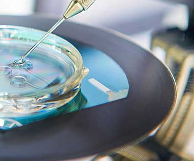
Embryo glue, sometimes referred to as an embryo transfer medium, is a specifically formulated fluid intended to resemble the uterus' organic environment. In the vital phase of implantation, it offers an adhesive property that aids the embryo in adhering to the uterine wall. The glue contains hyaluronan, a natural component in the female reproductive tract, which improves embryo-uterine lining interaction and attachment.

Embryo Glue Pros and Cons

Pros 

The ability to boost the implantation rate is one of the key benefits of employing embryo glue. According to studies, using embryo glue during IVF treatments may increase the likelihood of becoming pregnant. The embryo may stay in the desired position for implantation due to the glue's adhesive characteristics, which may lower the chance of embryo displacement during transfer. The glue may additionally improve the likelihood of successful implantation by facilitating an enhanced embryo-maternal connection.

Cons

Although embryo glue has the potential to increase implantation rates, scientific data supporting its efficacy is limited. Some studies document successful results, while others reveal no appreciable variation in conception rates from conventional transfer techniques. Embryo glue candidates must be aware of this limitation.

Like any medical procedure, glue components may cause allergic reactions or other side effects. Additionally, using embryo glue increases the cost of IVF procedures; this fact must be considered while designing a treatment plan.

Success Rate

Embryo glue may increase the likelihood of success by 7% to 9%. However, the efficacy rate of embryo glue can vary depending on patient characteristics and clinical expertise. Although some studies show that using embryo glue increases conception rates significantly, other research shows contradicting results. The efficacy of embryo glue may depend on the patient's age, health, and infertility history.

Side effects 

The side effects of embryo glue have been observed to be minor and similar to those associated with regular IVF techniques. Although serious adverse effects are extremely rare, some patients may feel little discomfort or irritability. 

Cost

The price of embryo glue varies based on the facility and area. It costs approximately Rs. 10,000/- to Rs. 20,000/- in India. The price range in the UK is between £200 and £400.

Alternatives

Even though embryo glue has demonstrated potential for improving implantation rates, it is not the sole choice. Different methods or processes, such as the use of a particular culture medium or embryo selection techniques, may be used by fertility clinics to improve embryo transfer. These consist of:

Natural Alternatives:

Uterine environment optimization

Endometrial scratching

Synthetic Substitutes:

Hyaluronic acid-based products

Synthetic adhesion molecules

Calcium-rich culture media

Conclusion

In a nutshell, embryo glue is a specific product designed to increase the likelihood of a fruitful embryo implantation during IVF procedures. Although it has the potential to increase implantation rates, there is currently insufficient scientific proof of its efficacy. Patients thinking about using embryo glue should speak with their fertility doctors, examine the benefits and drawbacks, and then decide depending on their individual situation and medical history.

FAQs

Is embryo glue safe to use during IVF?

While embryo glue is generally regarded as safe, people with allergies or other sensitivities should speak with their fertility doctors.

Does embryo glue guarantee a successful pregnancy?

No, embryo glue does not ensure pregnancy. It can, however, raise the likelihood of successful implantation.

How does embryo glue differ from traditional transfer methods?

The embryo's adhesion to the uterine wall during implantation is enhanced by the adhesive properties of embryo glue.

Can embryo glue be used in all IVF cases?

Embryo glue may be indicated in certain scenarios, however each patient's situation is unique and requires a specialized review.

What are the alternatives if I decide not to use embryo glue?

Other techniques or procedures for optimizing embryo transfer may be available at fertility clinics, and patients should discuss these options with their healthcare provider.

Equine Neurectomy Pros and Cons

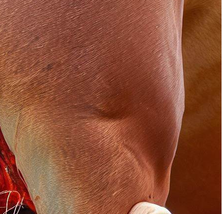
Equine neurectomy is a surgical treatment that involves cutting or removing a particular nerve, frequently the palmar/plantar digital nerves, in order to treat horses' persistent leg or hoof pain. This procedure disrupts the nerve's capacity to convey pain signals to the brain, thereby lowering or eliminating pain feelings in the affected area.

Equine Neurectomy Pros and Cons

The horse is given general anesthesia throughout the neurectomy process to ensure its safety and well-being. The veterinarian uses modern imaging tools to identify the targeted nerve and makes a tiny incision around the nerve site. Depending on the procedure, the nerve is then cut off or eliminated. The incision is carefully stitched up, and the horse is allowed time to recover from anesthesia.

Pros

Pain Relief

Equine neurectomy's ability to significantly reduce the pain experienced by horses with chronic limb or hoof pain is its main advantage. By blocking the pain impulses, the horse feels a significant reduction in discomfort, resulting in a better quality of life.

Enhancing Performance

Following a neurectomy, horses whose performance had previously been hampered by chronic pain frequently display improved performance. They are able to move with greater freedom and perform better in a variety of sports, including racing, jumping, and dressage, when they are pain-free.

Management of Chronic Conditions

Equine neurectomy is a viable alternative to conventional therapies in the management of chronic problems like laminitis or navicular syndrome. When all other measures have failed, it provides an alternate strategy for easing pain.

Cons

Loss of Feeling

The fact that neurectomy permanently destroys sensation in the affected area is a big problem. While this can help with pain management, it may also conceal injuries or other problems that the horse might have had in a normal situation.

Limited diagnostic abilities

Once the nerve is cut off it is more difficult to diagnose any potential problems that might develop later on in the treated area. Future treatment approaches may become more complicated as veterinarians struggle to identify new diseases or the cause of discomfort.

Risk of Overuse and Dependency

In some situations, neurectomy may be considered a quick remedy to a problem, leading to its misuse. Without treating the underlying source of the pain, relying entirely on this technique could result in dependency and future consequences.

Considering the Ethics

It is important to not take the decision to perform an equine neurectomy lightly. It raises moral concerns about the well-being of the horse and the procedure's possible long-term effects. Before making a final decision, horse owners and veterinarians should carefully assess whether neurectomy is the best option and look into alternative therapies.

Equine Neurectomy Alternatives

Horse owners and veterinarians should look into less invasive options before choosing neurectomy:

Medications

Without resorting to surgery, a number of drugs can assist manage pain and inflammation. Non-steroidal anti-inflammatory medicines (NSAIDs) and other painkillers may be effective and should be taken into consideration.

Rehabilitation and physical therapy

Physical therapy and rehabilitation can be incorporated into the horse's routine to assist recovery and naturally reduce pain. Controlled exercise, cold therapy, and heat therapy are all effective methods.

Alternative Therapies

Alternative pain management methods could be investigated as a complementary therapy, including acupuncture, chiropractic adjustments, and herbal supplements.

Upper Second Molar Extraction Pros and Cons

When a tooth poses a threat to oral health or cannot be preserved by other means, its extraction becomes necessary in dentistry. Upper second molar extraction is a frequent dental procedure carried out by dentists to treat various tooth disorders. 

Upper second molars are the teeth in the upper jaw that are located in the rear of the mouth. They play a key role in the overall oral structure by supporting neighboring teeth and aiding in chewing.

Upper Second Molar Extraction Pros and Cons

Reasons to Consider Extraction

Severe deterioration or damage

When an upper second molar is severely decayed or damaged, it may not be recoverable with fillings or root canal therapy.

Impacted Molars

Upper second molars can become impacted, which means they do not fully emerge or remain trapped behind the gum line. This may cause discomfort and even infections.

Crowding Issues

In rare situations, extracting an upper second molar may be necessary to solve crowding issues, particularly if there is insufficient space in the mouth for all of the teeth.

Periodontal Disease

Advanced periodontal disease can severely damage the supporting tissues of the upper second molars, necessitating extraction to avoid additional problems.

Pros of Upper Second Molar Extraction

Relief from Pain and Better Oral Health

A severely damaged or diseased upper second molar may need to be extracted for immediate pain relief. Additionally, it enhances general oral health by limiting the transmission of illness to neighboring tissues and teeth.

Avoiding Dental Problems

Potential dental concerns, like as misalignment and biting disorders, can be avoided by treating impacted molars or overcrowding during the extraction before they worsen.

Simpler Procedure

Upper second molar extraction is typically a simple procedure carried out by experienced dentists, lowering the risks associated with more complex dental procedures.

Creating Space for Orthodontic Treatment 

Removing the upper second molars might make the space needed to effectively align the remaining teeth in circumstances where orthodontic therapy is required.

Cons of Upper Second Molar Extraction

Affects Chewing Efficiency

Loss of an upper second molar might impair your ability to chew, especially when eating hard or rough meals.

Adjacent Teeth Susceptible to Movement

Following extraction, nearby teeth may gradually move in that direction, which could cause misalignment.

Dry Socket Risk

After an upper second molar extraction, a painful condition in which the blood clot at the extraction site dislodges early, might develop.

Need for Replacement Options

Following extraction, it can be important to look into tooth replacement options such dental implants or bridges to preserve optimal oral function and appearance.

FAQ's

Is upper second molar extraction painful?

Upper second molar extraction is carried out under local anesthetic to ensure little discomfort throughout the process. However, some post-operative tenderness may be felt; this can be treated with painkillers that the dentist will prescribe.

How long does it take to recover from an upper second molar extraction?

Although recovery times vary from person to person, the gum tissue typically takes about a week to completely recover. However, it can take a few weeks for the extraction site to fully recover.

Can I eat normally after the extraction?

Following the surgery, it's advised to eat only soft foods for a few days and refrain from chewing on the side of the extraction site. You can gradually start eating normally once the healing process is complete.

Will I need a replacement tooth after the extraction?

Your dentist will discuss tooth replacement possibilities with you based on your particular condition. Dental implants, bridges, or other options may be offered to maintain dental function and aesthetic.

Are there any alternatives to extracting an upper second molar?

A damaged tooth might occasionally be saved through dental procedures like root canal therapy. The best course of action, however, can be extraction if the tooth cannot be saved or if it compromises the overall health of the mouth.

Pec Implants Pros and Cons

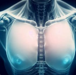
Pec implants are a type of cosmetic surgery used to improve the appearance of the chest muscles. Under the pectoral muscles, solid silicone implants are inserted during this treatment to give the chest a more defined, muscular appearance. Pec implants are a popular option for people who want a better-contoured chest but have struggled to obtain the required results with training and weightlifting alone.

Pec Implants Pros and Cons

Pros of Pec Implants

Improved Chest Appearance

The ability to obtain a more defined and sculpted chest is one of the main benefits of pec implants. People who are unable to naturally develop well-defined pectoral muscles due to heredity or other physical conditions may benefit from this therapy.

Boost in Self-Confidence

Many people equate masculinity and confidence with having a well-defined chest. Pec implants can provide a significant confidence boost by generating a more aesthetically pleasing chest contour, resulting in an enhanced body image and enhanced self-esteem.

symmetry and equilibrium

In order to improve the overall quality of life, a company must have a good deal of experience. Pec implants can help achieve a harmonious appearance by boosting the size and shape of the pectoral muscles, regardless of whether it is due to genetic causes or prior traumas.

Long-Term Approach

Pec implants provide a long-term solution for developing a contoured chest, in contrast to short-term solutions like chest padding or temporary fillers. Once the implants are in place, they remain in place and produce long-lasting effects.

Cons of Pec Implants

Risk of surgery

Pec implant surgery entails some dangers, just like any surgical procedure. These hazards include the potential for poor wound healing, infection, bleeding, and unfavorable anesthetic outcomes. 

Scarring

Incisions are made for insertion of the implants during Pec implant surgery, which may leave scars. However, expert surgeons use procedures to reduce scarring and ensure that incisions are placed in natural creases or hidden parts of the chest.

Recovery Time

Time and patience are needed for the recovery process following pec implant surgery. It's necessary to carefully adhere to the postoperative instructions, which may involve limiting physical activity and wearing compression clothing. The entire recovery process can take several weeks, and it is critical to give the body adequate time to heal properly.

Cost Considerations

The cost of the surgery is covered by the insurance company. The procedure's cost must be considered. The price of pec implants can fluctuate based on variables such as the skill of the surgeon, the location, facility fees, and anesthesia expenses.

Kybella Pros and Cons

A double chin, also known as submental fullness, can be reduced using the FDA-approved injectable procedure Kybella. It's become more well-known as a non-invasive substitute for chin liposuction. 

What is Submental Fullness?

Submental fullness is the buildup of fat beneath the chin, resulting in a double chin appearance. It can be caused by a variety of factors, including genetics, weight increase, and aging. While some people can reduce submental fullness by making lifestyle adjustments, others might need a more focused solution like Kybella.

What is Kybella?

Kybella is a prescription medication that contains synthetic deoxycholic acid, a naturally occurring chemical in the body that plays a role in the breakdown and absorption of dietary fat. Kybella works by destroying fat cells when injected into the submental region, which significantly reduces the appearance of submental fullness.

Kybella Pros and Cons

Pros

Efficient Submental Fat Loss

The efficiency of Kybella treatment in lowering submental fat is one of its primary advantages. When given by a licensed healthcare professional, clinical trials have demonstrated that Kybella can produce obvious contouring and a better chin profile.

Non-Surgical Technique

Kybella is a non-surgical alternative to conventional chin liposuction. It doesn't require incisions, anesthesia, or a protracted recovery period because it includes a series of injections directly into the fat behind the chin.

Minimal Downtime

The downtime following a Kybella injection is normally very short. After the procedure, some patients may feel mild swelling, bruising, or soreness, but these side effects typically go away within a few days, allowing people to resume their normal lives without too much disruption.

Long-Term Outcomes

The potential for long-lasting effects is another advantage of Kybella therapy. Once fat cells are destroyed, they can no longer accumulate or store fat. This suggests that the benefits obtained with Kybella can be sustained over time with a healthy lifestyle.

Enhances self-esteem and confidence

Submental fullness frequently causes people to feel self-conscious about their appearance. Enhancing chin contouring with Kybella therapy can aid with self-confidence and self-image.

Cons

Multiple Treatment Sessions

Multiple sessions separated by a few weeks are typically required for Kybella treatment. Although the precise number of sessions varies based on each patient's needs, the individual may need to be patient and dedicated to the treatment plan.

Bruising and swelling

Bruising and swelling are frequent side effects of Kybella therapy. Although these adverse effects are transient, anyone considering Kybella should be ready for some swelling and bruising during the healing process.

Cost Considerations

The price of Kybella treatment can be an issue. The overall cost could increase because numerous sessions are frequently necessary. A healthcare professional must be consulted in order to fully comprehend the total expenditure of therapy.

Not Suitable for Everyone

While Kybella is generally safe and effective, it is not appropriate for everyone. People should avoid receiving Kybella treatment if they have infections in the treatment area, blood disorders, or neck-related disorders. 

B-lite implants Pros and Cons

B-lite implants are a type of breast implant that uses cutting-edge technology to lighten the implant while preserving its strength and safety. These implants consist of a cohesive gel inside a silicone shell. The inclusion of microscopic hollow microspheres in the gel, which reduces the implant's overall weight, distinguishes them from the others.

B-lite implants Pros and Cons

Pros

Lightweight Design

The main benefit of B-lite implants is their considerably less weight. B-lite implants can be up to 30% lighter than conventional implants, making the procedure for the patient more comfortable. Because of the lessened stress on the breast tissues, there is less chance of long-term concerns like drooping or pain.

Reduced Strain on Tissues

B-lite implants more equally distribute the weight over the breast region because of their lighter makeup. The stress on the surrounding tissues is lessened by this equal distribution, potentially minimizing the chance of problems such implant malposition or capsular contracture. The lessened tension may also result in a more realistic-appearing and -feeling result.

Improved Comfort

In comparison to conventional implants, B-lite implants may provide more comfort. The likelihood of postoperative discomfort can be decreased because of the lighter weight, which can lessen the strain on the chest muscles. Since it allows for more freedom of movement and less constraint during physical activities, this feature is especially advantageous for people who lead an active lifestyle.

Enhanced Mobility

The reduced weight of B-lite implants enables more flexibility and mobility. Compared to conventional implants, patients may feel less restricted in their range of motion. This benefit can be especially important for athletes or people who regularly exercise because it makes it easier for them to engage in physical activity.

Cons

While B-lite implants have numerous advantages, some aspects should be examined before making a decision.

Cost

Because B-lite implants incorporate cutting-edge technology, they are typically more expensive than conventional implants.

Surgeon Expertise

Finding a surgeon with experience and knowledge of using B-lite implants is crucial because they are still a newer technology. 

Long-Term Resilience

Since B-lite implants are a more recent invention, research is currently being done to determine their long-term function and longevity. While preliminary evidence appears encouraging, it is critical to talk about the durability of B-lite implants with your surgeon.

Personal Preferences

Every person has different aspirations for their bodies and appearance. To ascertain whether B-lite implants are in line with your intended objectives and expectations, it is imperative to have a thorough discussion with your physician.

Limited Availability

It's possible that not all areas or clinics offer B-lite implants. Before considering them as a possibility, it's crucial to find out if these implants are offered where you live.

Conclusion

Potential advantages of B-lite implants include lighter weight, increased comfort, and increased mobility. Before selecting a choice, it's crucial to weigh aspects including cost, the skill of the surgeon, and long-term durability. You can decide if B-lite implants are the best option for your particular needs and goals by speaking with a trained plastic surgeon.

FAQs

Can B-lite implants rupture?

Yes, although there is a minimal likelihood of rupture, B-lite implants, like other breast implants, do carry a small risk of rupture.

Do B-lite implants require special care?

No, beyond the usual postoperative recommendations given by your surgeon, B-lite implants do not need any particular upkeep.

Are B-lite implants suitable for all body types?

The B-lite implants were made to fit a variety of body types. However, in order to choose the best solution for your unique physical traits, it's crucial to speak with a knowledgeable plastic surgeon.

Can B-lite implants be combined with other procedures?

It is possible to combine B-lite implants with additional surgeries like breast lifts or breast reductions. Your surgeon will evaluate your unique demands and advise you on the best course of action.

What is the lifespan of B-lite implants?

Depending on the individual, B-lite implants can last a lifetime. They can last for many years with the right maintenance and oversight.

Pros and Cons of Varithena

Varicose veins are bulging, twisted veins that frequently occur on the legs and can be uncomfortable as well as unpleasant. Varithena is a foam-based treatment that aims to lessen the discomfort of varicose veins and improve the overall appearance of the affected area. 

What is Varithena?

Varithena is a medical procedure that involves injecting a specially formulated foam containing the sclerosant agent polidocanol into the affected veins. The blood within the veins is displaced by this foam, which causes the veins to constrict and close. 

Pros and Cons of Varithena

Pros of Varithena

Non-Surgical Procedure

The fact that Varithena doesn't require surgery is one of its main benefits. Varithena does not need incisions or sutures, in contrast to conventional therapies such vein stripping or ligation. The foam is delivered directly into the problematic veins using a tiny catheter, causing the least amount of pain.

Minimally Invasive

Varithena is regarded as a minimally invasive treatment option for varicose veins. A tiny catheter is used to administer the foam, and it is normally placed through a very small puncture or incision. By using this method, the dangers of infection, scarring, and other open surgery-related consequences are avoided.

High Success Rate

Numerous studies have demonstrated the effectiveness of Varithena in treating varicose veins. The foam effectively seals off the injured veins, improving blood flow and providing symptom relief. Patients frequently report lessening of their discomfort, swelling, and pain as well as an improvement in the way the affected area looks.

Quick Recovery

Varithena offers a speedier recovery period than surgical procedures. After the therapy, the majority of patients can quickly return to their regular activities. Varithena's less invasive design shortens recovery time and enables faster return to normal activities.

Improved Aesthetic Appearance

Varithena can enhance the treated area's cosmetic appeal in addition to symptomatic relief. Varicose veins commonly appear as bulging, discolored veins on the skin's surface, which can be embarrassing for many people. Varithena assists in reducing the prominence of these veins, providing a more pleasing and natural appearance.

Cons of Varithena

Temporary Side Effects

Varithena, like any other medical procedure, may cause temporary adverse effects. These might include the treated area experiencing tightness, bruising, edema, or numbness. The majority of patients find these side effects manageable in light of the treatment's advantages because they normally go away within a few days or weeks.

Limited Availability

While Varithena has been increasingly popular in recent years, it may not be commonly available in all places. The treatment can only be performed in specific healthcare facilities since it needs specialized equipment and skilled medical personnel.

Cost Considerations

The price of Varithena is another factor to take into account. Varithena may be more expensive than more traditional management strategies like compression stockings. 

Varied Treatment Results

Although Varithena has a high percentage of success, individual treatment results may differ. Results may differ depending on a person's overall health, the severity of the illness, and the location of the affected veins. 

Potential Need for Multiple Sessions

Multiple Varithena treatment sessions can be required, depending on the severity of the varicose veins and the desired result. For adequate recovery and the best results, these treatments are often spaced apart by several weeks. During the initial consultation, you should talk to your healthcare physician about whether more sessions are necessary.

Conclusion

For those searching for a varicose veins treatment alternative, Varithena presents a potential choice. Many people find it to be an enticing option because to its non-surgical, minimally invasive nature, high success rate, short recovery, and aesthetic advantages. Before choosing a choice, it is crucial to take into account the possible short-term side effects, the availability constraints, the cost factors, the varied treatment outcomes, and the potential need for additional sessions.

Frequently Asked Questions (FAQs)

How long does a Varithena treatment session typically take?

Depending on the number and length of the veins that need to be treated with Varithena, a treatment session can last anywhere from 30 minutes to an hour.

Is Varithena painful?

Most patients say that the Varithena surgery caused them just minor discomfort. The use of local anesthetic helps to reduce any potential pain or discomfort.

Are the results of Varithena permanent?

Long-lasting improvements are possible with varithena, however new varicose veins could afterwards appear. Recurrence can be avoided by leading a healthy lifestyle and adhering to your doctor's advice.

Can Varithena be used for all types of varicose veins?

Varithena is appropriate for a variety of varicose vein types, however its suitability is dependent on the particular state of the veins. Your doctor will evaluate your condition and decide whether Varithena is the best course of treatment for you.

How soon can I expect to see results after Varithena treatment?

After having the Varithena procedure, patients frequently experience relief from their symptoms and an improvement in the overall appearance of their veins within a few weeks. Individual outcomes may vary.

Essentrics Pros and Cons

Physical fitness and well-being have grown to be important aspects of our lives, and many exercise techniques have gotten more and more popular over time. Essentrics is one technique that blends flexibility, strength, and stretching exercises. This article outlines Essentrics' pros and cons.

What is Essentrics?

The goal of the innovative exercise program Essentrics is to enhance body alignment, flexibility, and strength. Essentrics, a kind of exercise developed by former ballerina Miranda Esmonde-White, is influenced by a variety of practices, including Pilates, yoga, and ballet. It restores natural body movements and increases the functional range of motion.

Understanding the Essentrics Technique

Essentrics entails a mix of dynamic stretching and strengthening exercises carried out in a flowing, continuous way. The technique places a focus on eccentric muscular contractions, which involve extending the muscle while it is under tension. Essentrics uses these motions to rebalance the body, lengthen the muscles, and enhance posture.

Essentrics Pros and Cons

Pros of Essentrics

Improved flexibility and range of motion

Enhancing flexibility and range of motion is one of Essentrics' major advantages. Different muscle groups are targeted by the dynamic stretches done in Essentrics classes, which also aid in lengthening contracted muscles. Regular practice can promote joint mobility, making daily tasks easier and lowering the chance of injury.

Improved strength and muscular tone

By working for both major and small muscle groups, Essentrics offers a total-body workout. Resistance training combined with eccentric muscle contractions helps increase muscle tone and strength. Participants may observe body sculpting and muscular definition over time.

Relaxation and stress reduction

Exercises in Essentrics have advantages beyond the physical realm. The focused breathing and flowing motions induce meditation. This element of essencetrics has the potential to lessen stress, encourage relaxation, and enhance mental health in general.

Low-impact exercise for all fitness levels

Accessibility is one of the benefits of Essentrics. With less strain on the joints, it is a low-impact training technique. Essentrics is suited for persons of all fitness levels, including novices, elderly folks, and those recovering from injuries, in contrast to high-intensity workouts.

A multifaceted strategy for health and fitness

By focusing on the mind-body link, Essentrics adopts a holistic approach to health and fitness. To enhance general well-being, it integrates aspects of mindfulness, balance, and posture correction. After practicing Essentrics, many people say they feel more energized, balanced, and anchored.

Cons of Essentrics

Lack of cardiovascular benefits

Despite the fact that Essentrics has a lot of benefits, it's crucial to understand that its main emphasis is on body alignment, strength, and flexibility. Thus, it may not offer the same cardiovascular benefits as running or cycling. For those wishing to increase their cardiovascular fitness, Essentrics may need to be combined with extra cardio exercises.

Limited variety of exercises

The lack of variation in workouts offered by Essentrics in comparison to other fitness regimens may be another disadvantage. Despite the method's effectiveness, some people might want a more varied workout. To avoid boredom, combine Essentrics with other workout methods.

Risk of overstretching

Essentrics, like any fitness regimen, can cause damage if done incorrectly or pushed too far. To reduce the risk of overstretching or strain, it is essential to acquire the correct form and technique from an experienced instructor. Listening to the body and practicing within one's comfort zone are key for safe and efficient workouts.

Not suitable for some health conditions.

Essentrics is typically safe for most people, but not everyone should use it, especially if they have certain medical issues. Before starting Essentrics or any new exercise program, anyone with acute injuries, severe joint problems, or chronic diseases should visit a doctor.

FAQs

Is Essentrics suitable for weight loss?

Instead of emphasizing weight loss, Essentrics places a strong emphasis on body alignment, flexibility, and strength. But as part of a full fitness program, daily practice can help an active lifestyle and weight management.

How often should Essentrics be practiced?

Essentrics frequency depends on fitness and goals. Benefits often require 2-3 sessions per week. Long-term success requires consistency and regularity.

Can Essentrics help with posture improvement?

Yes, by strengthening the core muscles, extending short, tight muscles, and encouraging improved body alignment, Essentrics can aid in improving posture. Over time, good posture can be achieved with consistent practice and the right technique.

Is Essentrics similar to yoga or Pilates?

Although Essentrics has its own distinctive methodology, it draws inspiration from many disciplines such as yoga and Pilates. Essentrics focuses on eccentric muscular contractions and body balance, unlike yoga.

Are there any age restrictions for practicing Essentrics?

The truth is that Essentrics is appropriate for people of all ages. It includes low-impact workouts that prioritize joint safety and general well-being and may be tailored to different fitness levels, including older folks.

Sclerosing Epithelioid Fibrosarcoma Symptoms, Causes, Diagnosis, Treatment

What is Sclerosing Epithelioid Fibrosarcoma?

Sclerosing epithelioid fibrosarcoma (SEF) is a rare type of cancer that arises from fibrous connective tissue. It was first described in 1995 and has since been classified as a distinct type of fibrosarcoma due to its unique histological characteristics.

SEF is most commonly found in young adults, with a slight predilection for females. It usually arises in the deep soft tissues of the extremities, but can also occur in the trunk, head and neck region. SEF is a slow-growing tumor that tends to infiltrate surrounding tissues, making complete surgical excision challenging.

SEF is typically diagnosed through a combination of clinical examination, radiographic imaging, and histopathological analysis of a biopsy specimen. The tumor has a distinct appearance on imaging studies, often showing a solid, well-circumscribed mass with areas of calcification. Histologically, SEF is characterized by small, uniform, round cells arranged in nests or cords within a hyalinized stroma.

Sclerosing Epithelioid Fibrosarcoma Symptoms, Causes, Diagnosis, Treatment

Symptoms

The symptoms of sclerosing epithelioid fibrosarcoma (SEF) can vary depending on the location and size of the tumor. In some cases, SEF may not cause any symptoms until it grows to a size that compresses or invades nearby structures.

Common symptoms of SEF may include:

  • A painless lump or swelling in the affected area
  • Limited range of motion or stiffness in nearby joints
  • Numbness or tingling in the affected limb or body part
  • Weakness or loss of function in the affected limb or body part
  • Persistent pain that may worsen with activity or movement
  • Weight loss, fatigue, or other non-specific symptoms in advanced cases

Causes

The exact cause of sclerosing epithelioid fibrosarcoma (SEF) is not well understood. However, it is believed to arise from a genetic mutation or abnormality that leads to the uncontrolled growth of cells in fibrous connective tissue.

There is evidence to suggest that SEF may be associated with a genetic disorder called familial adenomatous polyposis (FAP), which is an inherited condition that increases the risk of developing polyps in the colon and rectum. Studies have found that individuals with FAP have an increased risk of developing SEF, particularly in the soft tissues of the extremities.

Other factors that may contribute to the development of SEF include exposure to radiation or certain chemicals, as well as trauma or injury to the affected area. However, the role of these factors in the development of SEF is not well established and more research is needed to understand the underlying causes of this rare cancer.

Diagnosis

Diagnosing sclerosing epithelioid fibrosarcoma (SEF) typically involves a combination of clinical examination, radiographic imaging, and histopathological analysis of a biopsy specimen.

The diagnostic process for SEF may involve the following steps:

Medical history and physical exam

A healthcare provider will review the patient's medical history and perform a physical exam to assess the location, size, and characteristics of the tumor.

Imaging tests

Imaging tests such as X-rays, CT scans, MRI scans, or PET scans may be used to visualize the tumor and evaluate its extent and location.

Biopsy

A biopsy is typically needed to confirm the diagnosis of SEF. During a biopsy, a small sample of the tumor tissue is removed and examined under a microscope by a pathologist to identify the presence of characteristic features of SEF.

Immunohistochemistry

Immunohistochemistry is a laboratory technique that uses specific antibodies to detect proteins or other markers in a tissue sample. It can help confirm the diagnosis of SEF and distinguish it from other types of tumors that may have similar characteristics.

Treatment

The treatment of sclerosing epithelioid fibrosarcoma (SEF) typically involves a multimodal approach that includes surgical excision, radiation therapy, and chemotherapy. The specific treatment plan will depend on factors such as the size, location, and extent of the tumor, as well as the patient's overall health and individual preferences.

Surgery

Surgery is the mainstay of treatment for SEF and involves the complete removal of the tumor. However, complete surgical excision of SEF can be challenging due to the tumor's tendency to infiltrate surrounding tissues. In some cases, amputation or limb-sparing surgery may be necessary to achieve complete removal of the tumor.

Radiation therapy

Radiation therapy may be used in combination with surgery to treat SEF. It involves the use of high-energy radiation to kill cancer cells and shrink the tumor. Radiation therapy may also be used as a palliative treatment to relieve pain and other symptoms in advanced cases.

Chemotherapy

Chemotherapy may be used to treat SEF that has spread to other parts of the body. However, SEF is typically resistant to chemotherapy, and its role in the treatment of this cancer is not well established.

Clinical trials

There is ongoing research into new treatments for SEF, and patients may be eligible to participate in clinical trials that test new drugs or therapies.

Scapulothoracic joint Function, Pain, Movements, Type

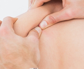
 The scapulothoracic joint is not a true joint in the traditional sense, as there is no bony articulation between the scapula (shoulder blade) and the thorax (rib cage). Instead, the scapula glides along the posterior thoracic wall, which is made up of the rib cage and associated musculature.

The scapulothoracic joint is an important component of normal shoulder motion, as it allows for a wide range of arm movements. The movement of the scapula is intimately linked with that of the shoulder joint proper (the glenohumeral joint), and the two joints must work together in order for normal shoulder function to occur.

Various muscles attach to the scapula and help to control its movement, including the trapezius, serratus anterior, and rhomboids. Dysfunction of these muscles can result in abnormal scapular movement and contribute to shoulder pain and dysfunction. Physical therapy and exercise programs are often used to address scapulothoracic dysfunction and improve shoulder function.

Scapulothoracic joint Function, Pain, Movements, Type

Function

The scapulothoracic joint plays an important role in shoulder movement and stability. Some of its key functions include:

Scapular rotation

The scapula rotates upward, downward, and laterally around the thorax to facilitate movement of the arm.

Scapular protraction/retraction

The scapula moves forward and backward along the thorax to enable arm movement and improve stability.

Scapular elevation/depression

The scapula moves up and down, helping to lift and lower the arm.

Scapular tilt

The scapula tilts forward and backward, which helps to maintain the proper alignment of the shoulder joint.

Stabilization

The scapula serves as a stable base for the shoulder joint, which allows the shoulder to move in a smooth and coordinated manner.

Pain

Scapular winging: This is a condition in which the scapula protrudes from the back due to weakness or paralysis of the serratus anterior muscle. This can cause pain and discomfort in the shoulder and upper back.

Shoulder impingement: This occurs when the tendons or bursa in the shoulder joint become compressed between the scapula and the humerus (upper arm bone). This can cause pain, weakness, and limited range of motion.

Thoracic outlet syndrome: This is a condition in which the nerves and blood vessels that pass through the thoracic outlet (the area between the neck and the shoulder) become compressed, causing pain, numbness, and tingling in the shoulder and arm.

Rotator cuff injury: The rotator cuff is a group of muscles and tendons that attach the scapula to the humerus. Injuries to the rotator cuff can cause pain and weakness in the shoulder and upper back.

Movements

The scapulothoracic joint allows for several movements that are important for shoulder function. Some of the main movements of the scapula include:

Upward rotation

This movement occurs when the inferior angle of the scapula moves away from the spine and upward, as in reaching overhead.

Downward rotation

This movement occurs when the inferior angle of the scapula moves toward the spine and downward, as in lowering the arm.

Protraction

This movement occurs when the scapula moves away from the spine and toward the front of the body, as in pushing or punching.

Retraction

This movement occurs when the scapula moves toward the spine and away from the front of the body, as in pulling or rowing.

Elevation

This movement occurs when the scapula moves upward toward the ear, as in shrugging.

Depression

This movement occurs when the scapula moves downward away from the ear, as in lowering the shoulders.

Type

The scapulothoracic joint is not a true synovial joint because it lacks the characteristics of a typical synovial joint, such as a joint capsule, articular cartilage, and a synovial membrane. Instead, the scapula glides along the posterior thoracic wall, which is made up of the rib cage and associated musculature.

The scapulothoracic joint is often classified as a "false joint" or a "pseudo-joint" because it is not a true joint in the traditional sense. However, it is still an important component of shoulder function and is intimately linked with the movement of the glenohumeral joint (shoulder joint proper).

Pedicle Subtraction Osteotomy Steps, Procedure, Recovery Time, Complications

What is Pedicle Subtraction Osteotomy?

Pedicle Subtraction Osteotomy (PSO) is a surgical procedure used to correct severe spinal deformities such as kyphosis, which is an excessive forward curvature of the spine. The PSO involves removing a wedge-shaped section of bone from the posterior aspect of the vertebral body, including the pedicle, lamina, and facet joints.

The goal of PSO is to realign the spine to a more normal curvature, which can alleviate symptoms such as back pain, difficulty standing or walking, and breathing problems. The procedure is typically performed on patients with severe deformities that have not responded to non-surgical treatments, such as bracing or physical therapy.

Steps

The specific steps involved in a Pedicle Subtraction Osteotomy (PSO) procedure can vary depending on the individual patient's condition and the surgeon's approach. However, some general steps typically involved in a PSO procedure may include:

Pedicle Subtraction Osteotomy Steps, Procedure, Recovery Time, Complications

Anesthesia

The patient is given general anesthesia, which will make them unconscious and unable to feel pain during the surgery.

Incision

The surgeon makes a long incision down the center of the back, exposing the spine.

Exposure

The surgeon removes the muscles and tissues covering the spinal column, exposing the affected vertebrae.

Pedicle removal

The surgeon removes the pedicle, which is a small bony protrusion on the back of the vertebrae, using specialized instruments.

Osteotomy

The surgeon then makes a precise cut through the vertebral body to create a wedge-shaped piece of bone, which is then removed. This allows the surgeon to change the angle of the spine and reduce the deformity.

Rod placement

The surgeon places metal rods along the length of the spine, connecting the vertebrae above and below the osteotomy site.

Bone grafting

The surgeon fills the space created by the osteotomy with a bone graft, which helps to stabilize the spine.

Closure

The surgeon closes the incision with sutures or staples and applies bandages.

Procedure

Pedicle Subtraction Osteotomy (PSO) procedure involves making a long incision down the center of the back, removing the muscles and tissues covering the spine, and removing a wedge-shaped section of bone from the posterior aspect of the affected vertebrae. The surgeon then places metal rods along the length of the spine and fills the space created by the osteotomy with a bone graft. The incision is closed with sutures or staples, and the patient will typically spend several days in the hospital for monitoring and pain management, followed by a rehabilitation program to regain strength and mobility.

Recovery

Recovery from a Pedicle Subtraction Osteotomy (PSO) can be a lengthy process and may vary from patient to patient. Generally, the patient can expect the following during the recovery process:

Hospital stay

The patient will spend several days in the hospital for monitoring and pain management.

Immobilization

The patient will need to wear a brace or cast for several weeks to immobilize the spine and allow the bone graft to heal.

Rehabilitation

Physical therapy and rehabilitation exercises will be necessary to help the patient regain strength and mobility in the affected area.

Pain management

The patient may experience pain and discomfort, which can be managed with medications prescribed by the surgeon.

Follow-up appointments

The patient will need to have follow-up appointments with the surgeon to monitor progress and make any necessary adjustments to the treatment plan.

Complications

As with any surgical procedure, Pedicle Subtraction Osteotomy (PSO) carries some risks and potential complications. Some of the most common complications associated with PSO may include:

Infection

There is a risk of infection with any surgical procedure, and patients undergoing PSO may develop an infection in the wound or in the bone graft site.

Nerve damage

PSO may involve manipulation of the spinal nerves, which can lead to nerve damage, including numbness, weakness, or paralysis.

Blood loss

PSO is a complex procedure that may involve significant blood loss, which can lead to anemia, shock, or other complications.

Anesthesia complications

General anesthesia carries a risk of complications, including reactions to the anesthesia, breathing difficulties, and other complications.

Failure to correct the deformity

In some cases, PSO may not successfully correct the spinal deformity or may lead to complications that require further surgical intervention.

Reed's Syndrome Symptoms, Causes, Testing, Treatment

What is Reed's Syndrome? 

Reed's syndrome, also known as lichen planus pemphigoides, is a rare autoimmune disorder that combines features of two different skin conditions: lichen planus and pemphigoid. Lichen planus is a condition that causes itchy, flat-topped, and purple-colored bumps on the skin or mucous membranes, while pemphigoid is a blistering skin disease that causes painful blisters and sores.

Reed's syndrome usually affects adults, and its symptoms can vary widely from person to person. Common symptoms of Reed's syndrome include itchy, reddish-purple bumps or plaques on the skin, blistering and erosions of the skin and mucous membranes, and nail abnormalities.

Reed's Syndrome Symptoms, Causes, Testing, Treatment

Reed's Syndrome Symptoms

The symptoms of Reed's syndrome can vary widely from person to person, but they generally involve a combination of skin and mucous membrane lesions. Some of the most common symptoms of Reed's syndrome include:

Reddish-purple bumps or plaques on the skin

These bumps or plaques may be flat or raised, and they can be itchy or painful. They often appear on the wrists, ankles, lower back, and the insides of the thighs.

Blisters and erosions

In some cases, Reed's syndrome can cause fluid-filled blisters on the skin or mucous membranes, which may break open and form painful erosions.

Nail abnormalities

Reed's syndrome can also affect the nails, causing ridges, grooves, or splitting.

Oral lesions

In some cases, Reed's syndrome can cause lesions on the inside of the mouth or other mucous membranes, leading to pain or difficulty eating or swallowing.

Itching

Many people with Reed's syndrome experience intense itching, which can be difficult to control.

Hair loss

In rare cases, Reed's syndrome may cause hair loss or thinning.

Reed's Syndrome Causes

The exact cause of Reed's syndrome is not yet fully understood, but it is believed to be an autoimmune disorder, which means that the body's immune system mistakenly attacks its own tissues. In Reed's syndrome, the immune system attacks the skin and mucous membranes, leading to the characteristic symptoms of the condition.

It is not clear what triggers the immune system to attack the body in this way, but several factors may be involved. These include genetic predisposition, exposure to certain medications or chemicals, and viral infections.

Some studies have suggested that Reed's syndrome may be associated with hepatitis C virus (HCV) infection, although not all people with Reed's syndrome have HCV, and not all people with HCV develop Reed's syndrome. Other viruses, such as the herpes simplex virus, have also been implicated in the development of Reed's syndrome.

In some cases, Reed's syndrome may occur as a reaction to medications or chemicals, such as certain antibiotics or dyes used in medical imaging tests. However, this is relatively rare.

Overall, the exact cause of Reed's syndrome is not yet fully understood, and further research is needed to better understand the underlying mechanisms and risk factors for this condition.

Reed's Syndrome Treatment

The treatment of Reed's syndrome depends on the severity of the symptoms and the individual patient's needs. In most cases, treatment involves a combination of medications and supportive care to manage symptoms and prevent complications.

Topical treatments

For mild cases of Reed's syndrome, topical medications such as corticosteroid creams or ointments may be used to reduce inflammation and itching.

Systemic medications

For more severe cases of Reed's syndrome, systemic medications may be necessary to suppress the immune system and reduce inflammation. These may include oral corticosteroids, immunosuppressant drugs, or biologic agents.

Phototherapy

Phototherapy, which involves exposing the skin to ultraviolet light, may be used to help reduce inflammation and itching in some cases of Reed's syndrome.

Supportive care

People with Reed's syndrome may need supportive care to manage symptoms such as itching, pain, and blistering. This may include taking cool baths or using cool compresses to soothe the skin, using gentle skin care products, and avoiding triggers such as certain medications or environmental factors that may worsen symptoms.

Dorsal scapular nerve Pain, Injury, Entrapment, Innervation

The dorsal scapular nerve is a nerve that arises from the cervical spinal nerves C5 and C6. It supplies the rhomboid muscles, which are responsible for retracting the scapula and elevating it. The nerve also innervates the levator scapulae muscle, which elevates the scapula and tilts the neck laterally.

The dorsal scapular nerve emerges from the posterior triangle of the neck and runs deep to the trapezius muscle. It is a motor nerve and does not have any sensory function. Damage to the dorsal scapular nerve can result in weakness or paralysis of the rhomboid and levator scapulae muscles, leading to shoulder and neck pain, limited shoulder movement, and shoulder blade winging.

Medical conditions that can cause dorsal scapular nerve damage include cervical spine injuries, cervical disc herniation, cervical spondylosis, and surgical injury. Treatment for dorsal scapular nerve injury typically involves physical therapy and exercises to improve muscle strength and range of motion. In severe cases, surgery may be necessary to repair nerve damage.

Dorsal scapular nerve Pain, Injury, Entrapment, Innervation

Dorsal scapular nerve Pain

The dorsal scapular nerve is a nerve that originates from the cervical spine and innervates the muscles that control the movement of the scapula, or shoulder blade. If this nerve becomes compressed or irritated, it can cause pain, weakness, and discomfort in the upper back, shoulder, and arm.

Common causes of dorsal scapular nerve pain include poor posture, repetitive strain injuries, trauma or injury to the neck or upper back, and cervical disc herniation. In some cases, nerve entrapment syndromes such as thoracic outlet syndrome or cervical rib syndrome may also be to blame.

Treatment for dorsal scapular nerve pain typically involves a combination of physical therapy, massage, and anti-inflammatory medication. In some cases, nerve blocks or surgical intervention may be necessary to relieve persistent pain or discomfort. If you are experiencing dorsal scapular nerve pain, it is important to consult with a healthcare professional to determine the underlying cause and develop an appropriate treatment plan.

Dorsal scapular nerve Injury

Dorsal scapular nerve injury occurs when there is damage or trauma to the dorsal scapular nerve, which controls the movement of the scapula or shoulder blade. The nerve originates from the cervical spine and travels through the upper back, where it innervates the rhomboid muscles and levator scapulae muscles.

Common causes of dorsal scapular nerve injury include direct trauma to the nerve, repetitive strain injuries, cervical spine surgery, or a neck injury such as whiplash. Symptoms of dorsal scapular nerve injury typically include pain, weakness, and decreased range of motion in the affected shoulder and upper back.

Dorsal scapular nerve Entrapment

Dorsal scapular nerve entrapment occurs when the dorsal scapular nerve becomes compressed or pinched along its course, leading to pain and dysfunction in the upper back and shoulder. The entrapment can occur at various locations, including at the level of the neck, where the nerve exits the cervical spine, or in the region of the rhomboid muscles, which the dorsal scapular nerve innervates.

Common causes of dorsal scapular nerve entrapment include poor posture, repetitive strain injuries, cervical disc herniation, or spinal stenosis. In some cases, underlying structural abnormalities such as a cervical rib or scoliosis can also contribute to nerve entrapment.

Symptoms of dorsal scapular nerve entrapment typically include pain, weakness, and decreased range of motion in the affected shoulder and upper back. Treatment options may include physical therapy, massage, and anti-inflammatory medication to alleviate pain and improve muscle function. In severe cases, nerve blocks or surgical intervention may be necessary to release the nerve from its entrapment.

Dorsal scapular nerve Innervation

The dorsal scapular nerve is a nerve that arises from the brachial plexus, specifically from the ventral rami of the C5 nerve root. It runs along the medial border of the scapula, and it innervates the rhomboid minor, rhomboid major, and the levator scapulae muscles.

The rhomboid minor muscle is responsible for retracting and elevating the scapula, while the rhomboid major muscle acts to retract and rotate the scapula. The levator scapulae muscle is responsible for elevating and rotating the scapula.

In addition to these muscles, the dorsal scapular nerve also sends branches to the trapezius muscle, which is responsible for elevating, retracting, and rotating the scapula. The dorsal scapular nerve also provides sensory innervation to the skin overlying the medial border of the scapula.

Sister Mary Joseph Nodule Symptoms, Causes, Life Expectancy, Treatment

Sister Mary Joseph Nodule?

Sister Mary Joseph nodule is a medical condition named after Sister Mary Joseph, who was a surgical assistant in the late 19th and early 20th centuries. The condition refers to the presence of a hard, palpable nodule or lump in the umbilical area, which can indicate the presence of an advanced cancer, such as stomach, ovarian, or pancreatic cancer.

The presence of a Sister Mary Joseph nodule often indicates that cancer has spread to the peritoneum, which is the thin layer of tissue that lines the abdominal cavity. This suggests that the cancer is advanced and may have already metastasized to other parts of the body.

The detection of a Sister Mary Joseph nodule is a significant finding and requires further diagnostic workup to determine the extent of the cancer and its treatment.

Sister Mary Joseph Nodule Symptoms, Causes, Life Expectancy, Treatment

Sister Mary Joseph Nodule Symptoms

Symptoms associated with a Sister Mary Joseph nodule include:

  • A hard, painful, and sometimes discolored nodule in the umbilical region
  • Swelling and redness around the nodule
  • Nausea and vomiting
  • Abdominal pain and discomfort
  • Unintentional weight loss
  • Anemia and fatigue
  • Change in bowel habits or blood in the stool
  • Abnormal vaginal bleeding or discharge

Sister Mary Joseph Nodule Causes

A Sister Mary Joseph nodule is not a primary disease but rather a secondary manifestation of an underlying malignancy. The nodule is a metastasis, which means that it is a site of cancer spread from a primary tumor located elsewhere in the body. The most common types of cancers associated with Sister Mary Joseph nodules are:

Gastrointestinal cancers

These include stomach, colon, pancreas, and liver cancers. These cancers spread to the umbilicus through the lymphatic system or by direct extension.

Gynecological cancers

These include ovarian, cervical, and uterine cancers. The spread to the umbilicus is through the peritoneal cavity and lymphatic channels.

Other less common cancers that can cause a Sister Mary Joseph nodule include lung cancer, breast cancer, and lymphoma.

The exact mechanism by which cancer cells reach the umbilicus is not fully understood. It is thought that cancer cells can spread through the lymphatic system, blood vessels, or by direct extension. The nodule can develop rapidly or slowly and may be the first sign of cancer or occur later in the disease course.

Sister Mary Joseph Nodule Life expectancy

Sister Mary Joseph nodule (SMJN) refers to a palpable nodule or mass that appears on the umbilicus and is a sign of an underlying malignancy, typically of the gastrointestinal or gynecological origin.

The life expectancy of an individual with SMJN depends on several factors, including the stage and type of cancer, the age and overall health of the patient, and the effectiveness of the treatments provided. In general, the presence of an SMJN is a sign of advanced cancer, which can reduce life expectancy.

Without treatment, the prognosis for an individual with SMJN is typically poor, with a median survival of only a few months. However, with appropriate treatment, including surgery, chemotherapy, and/or radiation therapy, the life expectancy of patients with SMJN can be improved, and some patients may even achieve long-term remission.

Sister Mary Joseph Nodule Treatment

The treatment of Sister Mary Joseph nodule (SMJN) depends on the underlying malignancy that is causing the nodule. SMJN is a sign of advanced cancer, and as such, the primary goal of treatment is typically palliative, to improve quality of life and prolong survival.

Treatment for SMJN may include surgery, chemotherapy, radiation therapy, or a combination of these approaches. The specific treatment plan will depend on several factors, including the type and stage of cancer, the location and size of the primary tumor, the extent of metastasis, and the overall health of the patient.

Surgery is often the first-line treatment for SMJN when feasible. The surgeon will typically remove the primary tumor and any affected lymph nodes and may also remove the nodule and surrounding tissue from the umbilicus.

Chemotherapy and radiation therapy may be used in combination with surgery or as a primary treatment for SMJN, depending on the type and stage of cancer. These treatments can help shrink the tumor and improve symptoms such as pain and bleeding.

What is HPV Cytopathic effect on women? - Definition, Assay, Examples

HPV, or human papillomavirus, is a common sexually transmitted virus that can cause abnormal growths or changes in the cells of the cervix, vagina, anus, or throat. When HPV infects the cells of the cervix, it can cause changes that are visible under a microscope. These changes are known as cytopathic effects, or CPE.

The cytopathic effects of HPV on women's cervical cells can vary from mild to severe. In mild cases, the changes may be minimal and may not require any treatment. However, in severe cases, the changes can lead to cervical cancer if left untreated.

HPV can cause several different types of CPE on the cervix, including:

Koilocytes

These are abnormal cells that have a characteristic "halo" or "raisin-like" appearance under the microscope. They are often associated with low-risk HPV types and are typically not cancerous.

Atypical squamous cells

These are abnormal cells that are not clearly cancerous, but are not entirely normal either. They may be caused by both low-risk and high-risk HPV types.

Dysplasia

This refers to the abnormal growth of cells on the surface of the cervix. It is typically caused by high-risk HPV types and can progress to cancer if left untreated.

What is HPV Cytopathic effect on women - Definition, Assay, Examples

Definition

Cytopathic effect, or CPE, is the observable changes that occur in the appearance, behavior, or function of cells in response to a viral infection or other pathogenic agent. The changes may include cell rounding, detachment, fusion, or the formation of syncytia, which are multinucleated cells resulting from the fusion of infected and uninfected cells.

The cytopathic effect can be caused by the direct effects of viral replication or by the host immune response to the infection. In some cases, the cytopathic effect can lead to cell death or tissue damage, which can contribute to the pathogenesis of the infection.

The cytopathic effect can be observed using various techniques, including light microscopy, electron microscopy, or fluorescence microscopy. It is an important tool for diagnosing and studying viral infections, as well as for evaluating the efficacy of antiviral drugs or vaccines.

Assay

Cytopathic effect assay, also known as CPE assay, is a laboratory technique used to identify and quantify the effects of a virus on cultured cells. The assay involves infecting a cell culture with a virus and monitoring the resulting changes in the appearance and behavior of the cells.

In the case of HPV, the cytopathic effect assay can be used to assess the effect of the virus on cervical cells in vitro. The assay can detect changes in the morphology and viability of the cells, as well as the presence of specific markers of HPV infection.

The cytopathic effect assay is a useful tool for studying the behavior and virulence of viruses in a controlled laboratory environment. It can also be used to evaluate the efficacy of antiviral drugs or vaccines, as well as to screen for new antiviral agents.

However, it is important to note that the results of the cytopathic effect assay may not always be representative of the effects of the virus in vivo, and additional tests and studies may be needed to fully understand the clinical implications of HPV infection.

Examples

Some examples of cytopathic effects caused by viral infections include:

  • Herpes simplex virus (HSV) infection can cause the formation of multinucleated giant cells and cellular destruction in infected cells.
  • Cytomegalovirus (CMV) infection can cause enlarged cells with an increased number of nuclei, called "owl's eye" cells.
  • Influenza virus infection can cause cell rounding, detachment, and formation of syncytia.
  • Human papillomavirus (HPV) infection can cause the appearance of abnormal cells, such as koilocytes or atypical squamous cells, or the development of dysplasia, which is the abnormal growth of cells on the surface of the cervix.
  • Measles virus infection can cause giant cell formation and syncytia in respiratory epithelial cells.

Mauriac Syndrome Symptoms, Causes, Diagnosis, Edema, Treatment

Mauriac syndrome, also known as Malnutrition-associated diabetes mellitus, is a rare complication of poorly controlled diabetes mellitus in children. This condition was first described by the French physician François Mauriac in the 1930s.

Mauriac syndrome is characterized by stunted growth, delayed puberty, and a distinctive body habitus, which includes abdominal distension, thin extremities, and a moon-shaped face. These physical changes are a result of malnutrition, which occurs because insulin therapy in poorly controlled diabetes can lead to decreased appetite and increased breakdown of fats and proteins in the body.

Symptoms

The symptoms of Mauriac syndrome, also known as malnutrition-associated diabetes mellitus, may include:

Stunted growth

Children with Mauriac syndrome often have delayed growth and are smaller than their peers.

Delayed puberty

Puberty may be delayed or absent in children with Mauriac syndrome.

Moon-shaped face

Children with Mauriac syndrome may have a round, moon-shaped face due to excess fat deposits.

Thin extremities

The arms and legs may appear thin compared to the torso due to the breakdown of fats and proteins in the body.

Abdominal distension

The abdomen may appear swollen or distended due to the buildup of fluid in the abdominal cavity.

Poor appetite

Children with Mauriac syndrome may have a decreased appetite due to high blood sugar levels.

Fatigue

High blood sugar levels can also lead to fatigue and lethargy

Mauriac Syndrome Symptoms, Causes, Diagnosis, Edema, Treatment

Causes

Mauriac syndrome, also known as malnutrition-associated diabetes mellitus, is caused by prolonged poorly controlled diabetes in children. When blood sugar levels are not well-managed, it can lead to a range of complications, including malnutrition.

The underlying mechanism of Mauriac syndrome involves insulin resistance, which is a common feature of poorly controlled diabetes. Insulin resistance makes it difficult for cells to use glucose for energy, leading to high levels of glucose in the bloodstream. This, in turn, can cause decreased appetite, increased fat breakdown, and muscle wasting, which can contribute to malnutrition.

Additionally, high blood sugar levels can also lead to the accumulation of fluid in the abdomen, a condition known as ascites. Ascites can cause abdominal distension and further contribute to the malnourished appearance seen in children with Mauriac syndrome.

Diagnosis

The diagnosis of Mauriac syndrome, also known as malnutrition-associated diabetes mellitus, typically involves a combination of clinical evaluation, blood tests, and imaging studies.

Clinical evaluation

A doctor may perform a physical exam to assess the child's growth and development, look for signs of malnutrition, such as thin extremities, abdominal distension, and a moon-shaped face, and assess the child's pubertal development.

Blood tests

Blood tests may be done to evaluate blood sugar levels and other markers of glycemic control, such as glycated hemoglobin (HbA1c) and fasting blood glucose levels. Blood tests may also be done to assess for malnutrition, such as albumin and prealbumin levels.

Imaging studies

Imaging studies, such as an abdominal ultrasound or CT scan, may be done to evaluate for the presence of ascites, which can contribute to abdominal distension.

Edema

Mauriac syndrome, also known as malnutrition-associated diabetes mellitus, can lead to the development of edema in some cases. The accumulation of fluid in the abdominal cavity, known as ascites, is a common complication of poorly controlled diabetes and can cause abdominal distension and edema in the legs and feet.

The underlying mechanism of edema in Mauriac syndrome is multifactorial. Prolonged high blood sugar levels can lead to damage to the blood vessels, making it difficult for fluid to be transported efficiently from the tissues to the blood vessels. Additionally, protein loss due to malnutrition can also contribute to edema, as proteins help to maintain the balance of fluid in the tissues.

Treatment

The treatment of Mauriac syndrome, also known as malnutrition-associated diabetes mellitus, typically involves a multifaceted approach that includes optimizing glycemic control, nutritional support, and treatment of associated complications.

Glycemic control

The cornerstone of treatment for Mauriac syndrome is achieving and maintaining good glycemic control. This may involve intensive insulin therapy, including the use of multiple daily injections or insulin pump therapy, to regulate blood sugar levels.

Nutritional support

Children with Mauriac syndrome are often malnourished and require nutritional support to improve their overall health and well-being. This may include a balanced diet rich in protein, carbohydrates, and healthy fats, as well as vitamin and mineral supplementation.

Treatment of complications

Complications associated with Mauriac syndrome, such as edema and delayed growth and development, may require additional treatment. This may include the use of diuretics to reduce fluid buildup, growth hormone therapy to improve growth, and surgery to address complications such as ascites.

Osteoclastogenesis Definition, Process, Pathway, Inhibitory factors

Osteoclastogenesis is the process by which osteoclasts, the bone-resorbing cells, are formed from hematopoietic stem cells (HSCs) in bone marrow. Osteoclasts are essential for bone remodeling, which is the process by which old or damaged bone tissue is removed and replaced by new bone tissue.

The process of osteoclastogenesis is regulated by several factors, including the receptor activator of nuclear factor kappa B ligand (RANKL), macrophage colony-stimulating factor (M-CSF), and osteoprotegerin (OPG). RANKL is produced by osteoblasts and binds to its receptor RANK on the surface of osteoclast precursor cells, promoting their differentiation into mature osteoclasts. M-CSF stimulates the proliferation of osteoclast precursor cells, and OPG acts as a decoy receptor for RANKL, preventing it from binding to RANK and inhibiting osteoclastogenesis.

Osteoclastogenesis plays a critical role in bone development, growth, and maintenance, as well as in the pathogenesis of various bone diseases, including osteoporosis, rheumatoid arthritis, and bone metastases. Therefore, understanding the mechanisms underlying osteoclastogenesis is essential for the development of new treatments for these conditions.

Definition

Osteoclastogenesis is the process by which osteoclasts, the bone-resorbing cells, are formed from hematopoietic stem cells (HSCs) in bone marrow. It involves a series of steps, including the differentiation of osteoclast precursor cells into mature osteoclasts, the fusion of these cells to form multinucleated osteoclasts, and the activation of these cells to resorb bone tissue. 

Osteoclastogenesis Definition, Process, Pathway, Inhibitory factors

Process

Osteoclastogenesis is a complex process that involves several steps. Here is a simplified overview of the process:

  • Hematopoietic stem cells (HSCs) differentiate into osteoclast precursor cells in the bone marrow.
  • The osteoclast precursor cells are then stimulated by macrophage colony-stimulating factor (M-CSF), which promotes their proliferation and survival.
  • The receptor activator of nuclear factor kappa B ligand (RANKL), produced by osteoblasts and other cells, binds to its receptor RANK on the surface of osteoclast precursor cells, triggering a signaling cascade that promotes their differentiation into mature osteoclasts.
  • The osteoclast precursor cells fuse together to form multinucleated osteoclasts.
  • The mature osteoclasts are then activated to resorb bone tissue by secreting enzymes and acids that break down the mineral and organic components of bone.
  • Osteoprotegerin (OPG), produced by osteoblasts and other cells, can bind to RANKL and prevent it from binding to RANK, thus inhibiting osteoclastogenesis.

Pathway

The osteoclastogenesis pathway involves several signaling molecules and transcription factors that regulate the differentiation of osteoclast precursor cells into mature osteoclasts. Here is a simplified overview of the pathway:

  1. Hematopoietic stem cells differentiate into osteoclast precursor cells in the bone marrow.
  2. Osteoclast precursor cells express the receptor activator of nuclear factor kappa B (RANK) on their surface.
  3. The binding of RANKL to RANK on osteoclast precursor cells triggers a signaling cascade that activates several transcription factors, including nuclear factor of activated T cells cytoplasmic 1 (NFATc1), activator protein 1 (AP-1), and nuclear factor kappa B (NF-κB).
  4. NFATc1 is a key transcription factor that induces the expression of genes involved in osteoclast differentiation, including tartrate-resistant acid phosphatase (TRAP), cathepsin K, and matrix metalloproteinase 9 (MMP-9).
  5. AP-1 and NF-κB also play important roles in regulating osteoclastogenesis by inducing the expression of genes involved in osteoclast differentiation and survival.
  6. The activation of NFATc1, AP-1, and NF-κB is regulated by various signaling pathways, including the mitogen-activated protein kinase (MAPK) pathway, the phosphoinositide 3-kinase (PI3K) pathway, and the protein kinase C (PKC) pathway.
  7. Osteoprotegerin (OPG) acts as a decoy receptor for RANKL, preventing it from binding to RANK and inhibiting osteoclastogenesis.

Inhibitory Factors

Several factors can inhibit osteoclastogenesis and bone resorption. Here are some examples:

Osteoprotegerin (OPG)

OPG is a decoy receptor for RANKL, which prevents RANKL from binding to RANK on the surface of osteoclast precursor cells, thus inhibiting osteoclastogenesis.

Interleukin-4 (IL-4)

IL-4 is a cytokine that inhibits osteoclastogenesis by blocking the differentiation of osteoclast precursor cells into mature osteoclasts.

Interleukin-10 (IL-10)

IL-10 is an anti-inflammatory cytokine that inhibits osteoclastogenesis by downregulating the expression of RANK on osteoclast precursor cells.

Interferon gamma (IFN-γ)

IFN-γ is a cytokine that inhibits osteoclastogenesis by suppressing the expression of RANKL and M-CSF by osteoblasts and other cells.

Bisphosphonates

Bisphosphonates are drugs that inhibit bone resorption by targeting osteoclasts and inducing their apoptosis.

Estrogen

Estrogen is a hormone that inhibits osteoclastogenesis by decreasing the expression of RANKL and increasing the expression of OPG.

Trending Topics

Powered by Blogger.
Scroll To Top