Osteoclastogenesis Definition, Process, Pathway, Inhibitory factors

Osteoclastogenesis is the process by which osteoclasts, the bone-resorbing cells, are formed from hematopoietic stem cells (HSCs) in bone marrow. Osteoclasts are essential for bone remodeling, which is the process by which old or damaged bone tissue is removed and replaced by new bone tissue.

The process of osteoclastogenesis is regulated by several factors, including the receptor activator of nuclear factor kappa B ligand (RANKL), macrophage colony-stimulating factor (M-CSF), and osteoprotegerin (OPG). RANKL is produced by osteoblasts and binds to its receptor RANK on the surface of osteoclast precursor cells, promoting their differentiation into mature osteoclasts. M-CSF stimulates the proliferation of osteoclast precursor cells, and OPG acts as a decoy receptor for RANKL, preventing it from binding to RANK and inhibiting osteoclastogenesis.

Osteoclastogenesis plays a critical role in bone development, growth, and maintenance, as well as in the pathogenesis of various bone diseases, including osteoporosis, rheumatoid arthritis, and bone metastases. Therefore, understanding the mechanisms underlying osteoclastogenesis is essential for the development of new treatments for these conditions.

Definition

Osteoclastogenesis is the process by which osteoclasts, the bone-resorbing cells, are formed from hematopoietic stem cells (HSCs) in bone marrow. It involves a series of steps, including the differentiation of osteoclast precursor cells into mature osteoclasts, the fusion of these cells to form multinucleated osteoclasts, and the activation of these cells to resorb bone tissue. 

Osteoclastogenesis Definition, Process, Pathway, Inhibitory factors

Process

Osteoclastogenesis is a complex process that involves several steps. Here is a simplified overview of the process:

  • Hematopoietic stem cells (HSCs) differentiate into osteoclast precursor cells in the bone marrow.
  • The osteoclast precursor cells are then stimulated by macrophage colony-stimulating factor (M-CSF), which promotes their proliferation and survival.
  • The receptor activator of nuclear factor kappa B ligand (RANKL), produced by osteoblasts and other cells, binds to its receptor RANK on the surface of osteoclast precursor cells, triggering a signaling cascade that promotes their differentiation into mature osteoclasts.
  • The osteoclast precursor cells fuse together to form multinucleated osteoclasts.
  • The mature osteoclasts are then activated to resorb bone tissue by secreting enzymes and acids that break down the mineral and organic components of bone.
  • Osteoprotegerin (OPG), produced by osteoblasts and other cells, can bind to RANKL and prevent it from binding to RANK, thus inhibiting osteoclastogenesis.

Pathway

The osteoclastogenesis pathway involves several signaling molecules and transcription factors that regulate the differentiation of osteoclast precursor cells into mature osteoclasts. Here is a simplified overview of the pathway:

  1. Hematopoietic stem cells differentiate into osteoclast precursor cells in the bone marrow.
  2. Osteoclast precursor cells express the receptor activator of nuclear factor kappa B (RANK) on their surface.
  3. The binding of RANKL to RANK on osteoclast precursor cells triggers a signaling cascade that activates several transcription factors, including nuclear factor of activated T cells cytoplasmic 1 (NFATc1), activator protein 1 (AP-1), and nuclear factor kappa B (NF-κB).
  4. NFATc1 is a key transcription factor that induces the expression of genes involved in osteoclast differentiation, including tartrate-resistant acid phosphatase (TRAP), cathepsin K, and matrix metalloproteinase 9 (MMP-9).
  5. AP-1 and NF-κB also play important roles in regulating osteoclastogenesis by inducing the expression of genes involved in osteoclast differentiation and survival.
  6. The activation of NFATc1, AP-1, and NF-κB is regulated by various signaling pathways, including the mitogen-activated protein kinase (MAPK) pathway, the phosphoinositide 3-kinase (PI3K) pathway, and the protein kinase C (PKC) pathway.
  7. Osteoprotegerin (OPG) acts as a decoy receptor for RANKL, preventing it from binding to RANK and inhibiting osteoclastogenesis.

Inhibitory Factors

Several factors can inhibit osteoclastogenesis and bone resorption. Here are some examples:

Osteoprotegerin (OPG)

OPG is a decoy receptor for RANKL, which prevents RANKL from binding to RANK on the surface of osteoclast precursor cells, thus inhibiting osteoclastogenesis.

Interleukin-4 (IL-4)

IL-4 is a cytokine that inhibits osteoclastogenesis by blocking the differentiation of osteoclast precursor cells into mature osteoclasts.

Interleukin-10 (IL-10)

IL-10 is an anti-inflammatory cytokine that inhibits osteoclastogenesis by downregulating the expression of RANK on osteoclast precursor cells.

Interferon gamma (IFN-γ)

IFN-γ is a cytokine that inhibits osteoclastogenesis by suppressing the expression of RANKL and M-CSF by osteoblasts and other cells.

Bisphosphonates

Bisphosphonates are drugs that inhibit bone resorption by targeting osteoclasts and inducing their apoptosis.

Estrogen

Estrogen is a hormone that inhibits osteoclastogenesis by decreasing the expression of RANKL and increasing the expression of OPG.

Right & Left Internal Carotid Artery Occlusion Symptoms, Causes, Treatment

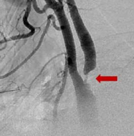
Carotid artery occlusion refers to the blockage or narrowing of the carotid artery, which is one of the major arteries supplying blood to the brain. This condition is often caused by atherosclerosis, a buildup of plaque in the artery walls, which can lead to the formation of a blood clot that completely blocks blood flow.

Right & Left internal carotid artery occlusion

Internal carotid artery occlusion is a serious medical condition that can cause significant morbidity and mortality. The internal carotid arteries are two of the major arteries that supply blood to the brain, and their occlusion can lead to a reduction or complete loss of blood flow to the affected side of the brain.

When both the right and left internal carotid arteries are occluded, it is called bilateral internal carotid artery occlusion. This is a rare condition but can lead to severe neurological deficits and disability.

The symptoms of bilateral internal carotid artery occlusion can include a sudden loss of consciousness, seizure, or stroke-like symptoms such as weakness or numbness on one side of the body, difficulty speaking, or vision changes.

Right & Left internal carotid artery occlusion Symptoms, Causes, Treatment

Symptoms

The symptoms of internal carotid artery occlusion can vary depending on the severity and location of the blockage. The following are some of the common symptoms:

  • Sudden onset of weakness or numbness on one side of the body, face, or limbs.
  • Difficulty speaking or understanding speech.
  • Vision changes, such as double vision or partial or complete loss of vision in one eye.
  • Sudden, severe headache, especially in the absence of a known cause.
  • Dizziness or loss of balance.
  • Difficulty with coordination or walking.
  • Confusion, disorientation, or memory loss.
  • Loss of consciousness, seizure, or coma.

Causes

Internal carotid artery occlusion is most commonly caused by atherosclerosis, which is the buildup of plaque in the walls of the arteries that can lead to narrowing or complete blockage of the artery. Other possible causes of internal carotid artery occlusion include:

  • Blood clots or emboli that form in other parts of the body and travel to the brain, blocking the internal carotid artery.
  • Inflammation or infection of the artery, such as in the case of temporal arteritis.
  • Trauma or injury to the artery, such as a carotid artery dissection.
  • Congenital abnormalities of the artery, such as fibromuscular dysplasia or arterial tortuosity syndrome.
  • Rare genetic conditions that affect the structure or function of the artery, such as Ehlers-Danlos syndrome.
  • Radiation therapy to the neck or head, which can cause scarring and narrowing of the artery.

Treatment

The treatment for internal carotid artery occlusion depends on the severity of the blockage and the symptoms that the patient is experiencing. The goal of treatment is to prevent further blockage or complications and to improve blood flow to the affected area of the brain. Here are some common treatment options:

Medications
Blood thinners or antiplatelet drugs such as aspirin, clopidogrel, or warfarin may be prescribed to prevent blood clots from forming and reduce the risk of stroke.

Lifestyle modifications
Quitting smoking, losing weight, and managing blood pressure and cholesterol through diet and exercise can help improve overall cardiovascular health and reduce the risk of further blockage.

Carotid endarterectomy
This is a surgical procedure in which a surgeon removes the plaque buildup from the inner lining of the carotid artery to restore blood flow to the brain.

Carotid angioplasty and stenting
This is a less invasive procedure that involves the insertion of a catheter with a balloon into the blocked artery to widen it and a stent to keep it open.

Extracranial-intracranial bypass surgery
This is a surgical procedure in which a surgeon connects a blood vessel from outside the skull to a blood vessel inside the skull to bypass the blocked artery and improve blood flow to the brain.

Trending Topics

Powered by Blogger.
Scroll To Top